This article provides a protocol for the systematic approach to the technique of Mohs micrographic surgery. Each step, from tumor excision and tissue mapping, to specimen processing and histologic interpretation, through wound closure and postoperative management, is covered. The advantages of Mohs surgery over other treatment modalities are observed histologic margin control, superior cure rates, and maximal tissue-sparing potential. The increased preservation of normal tissue leads to smaller surgical defects, optimal reconstructive results, and diminished risk of poor surgical outcomes. Overall, the risks of the procedure are few, the benefits numerous, and the outcomes worth the time and effort spent in learning the technique.
Mohs micrographic surgery (MMS) consists of a standardized series of steps in cutaneous surgery for the purpose of extirpation of skin cancers. The advantages of MMS over standard excision are precise and thorough histologic margin control, superior cure rates, and maximal preservation of normal tissue. It is a technique developed for the management of cutaneous tumors that grow in predictably contiguous fashion, such that once the excision has been observed histologically to be carried out beyond the boundary of the tumor, a surgeon can feel confident that a true negative margin has been achieved. This fact is reflected in the superior cure rates of MMS compared with traditional surgical excision. The beauty of the technique lies in the simplicity of its individual steps, but the elegance and synergy of those steps in aggregate. Furthermore, when MMS is the chosen method for tumor clearance in accordance with the generally accepted indications for the procedure, the average cost to patients and the health care system as a whole is at least equal to, if not less than, traditional surgical excision. This article provides a protocol for the systematic approach to skin cancer excision and tissue mapping, transportation and processing of the surgical specimen, histopathology interpretation, and wound closure and management.
Indications for Mohs micrographic surgery
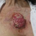
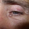
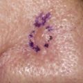
Prior to performing MMS on a given tumor, it must be determined if the technique is indicated for the particular lesion in question. There is general acceptance of most indications for MMS, which are based on tumor size and histology as well as anatomic location and previous treatments ( Box 1 and Fig. 1 ). As the discipline of MMS continues to evolve, new applications for the technique draw support and criticism. There have been attempts to utilize MMS in the treatment of other types of skin cancer that have traditionally been excised with wide local excision. There are reports of utilizing MMS for lentigo maligna melanoma, invasive malignant melanoma, Merkel cell carcinoma, dermatofibrosarcoma protuberans, sebaceous carcinoma, extramammary Paget disease, and microcystic adnexal carcinoma, among other skin cancers, with varying success rates. Some investigators now consider these tumor types to be reasonable indications for MMS.
Nonmelanoma skin cancers (basal cell carcinoma and squamous cell carcinoma) greater than 0.4 cm in high-risk location (H zone of face)
Large tumors (>1 cm on the face; >2 cm on the trunk and extremities)
Recurrent and/or incompletely excised tumors
Tumors with aggressive histologic subtypes or indistinct clinical borders (infiltrative, micronodular, and morpheaform basal cell carcinoma; basal cell carcinoma and squamous cell carcinoma with perineural or perivascular invasion)
Tumors in cosmetically sensitive or functionally important locations (genital, anal, hand, and foot locations)
Nonmelanoma skin cancer arising in an immunosuppressed patient
Tumors arising in sites of chronic inflammation, long-standing wounds, burns, or scars
Genetic conditions predisposing to many skin cancers (eg, basal cell nevus syndrome, xeroderma pigmentosa, and Muir-Torre syndrome)
Related posts:
 Mohs Surgery for Squamous Cell Carcinoma
 An Overview of Mohs Micrographic Surgery for the Treatment of Basal Cell Carcinoma
 Management of Unusual Cutaneous Malignancies: Atypical Fibroxanthoma, Malignant Fibrous Histiocytoma, Sebaceous Carcinoma, Extramammary Paget Disease
 Management of Skin Cancer in Solid-organ Transplant Recipients: A Multidisciplinary Approach
Special Considerations for Mohs Micrographic Surgery on the Eyelids, Lips, Genitalia, and Nail Unit
Multidisciplinary Approach to Large Cutaneous Tumors of the Head and Neck
Management of Skin Cancer in Solid-organ Transplant Recipients: A Multidisciplinary Approach
Special Considerations for Mohs Micrographic Surgery on the Eyelids, Lips, Genitalia, and Nail Unit
Multidisciplinary Approach to Large Cutaneous Tumors of the Head and Neck
Stay updated, free articles. Join our Telegram channel

Full access? Get Clinical Tree


