Fig. 26.1
Patients with chronic bilateral advanced lymphedema
Campisi et al. described successful outcomes of micro-lymphaticovenous anastomoses (LVA) for moderate-stage lymphedema. However, this procedure is not favored for Campisi stage IV (fibrotic and column-shaped limb) and Campisi stage V (elephantiasis with severe limb deformation, scleroindurative pachidermatitis and widespread lymphstatic verrucosis) advanced lymphoedema. They also support microlymphatic surgery only for the obstructive type of lymphoedema characterized by lymphadenodysplasia, but not for lymphatic-lymphonodal aplasia [9, 10]. On the other hand, Koshima et al. have recommended microsurgical lymphaticovenous anastomoses for primary and secondary lymphoedema, even in advanced stages IV and V. They base this recommendation on the ultrastructural observations of human lymphatics in edematous limbs [11, 12].
We, however, have not been successful in performing lymphaticovenous anastomoses in advanced lymphoedema and we think this is because the lymphatic trunks are neither patent nor functional. Recurrent episodes of infection and cellulitis lead to subcutaneous fibrosis, thereby occluding any remaining functional lymphatic channels. A vicious cycle develops in which the edema worsens, causing further damage to the subcutaneous tissue, rendering it unreliable for LVA. In anaplastic and hypoplastic types of primary lymphoedema, there are only a few or no lymphatics. Microsurgery, therefore, has little role to play in the treatment of these patients. Infection, with recurrent cellulitis and lymphangitis, is an undesirable complication and upgrades the stage of the disease, adding to the patient’s frustration and lack of well-being. Control of infection is therefore paramount in advanced disease. The excisional technique of the Charles’ procedure can produce good long-term results and is a realistic approach in advanced lymphedema [3, 7].
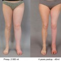
Recent evidence suggests that the Charles’ procedure can result in a 30 % reduction of the circumference, which can be maintained over a 3-year period, resulting in a 90 % reduction in cellulitis [3].
Potential complications of the procedure are wound breakdown, hyperkeratosis, ulceration, and aggravation of foot lymphedema (Fig. 26.2). To prevent these problems many authors described a modified Charles’ procedure. Van der Walt et al. presented a modified Charles’ procedure, applying negative-dressing after the initial debulking surgery and then they delayed the skin grafting by 5–7 days [13].
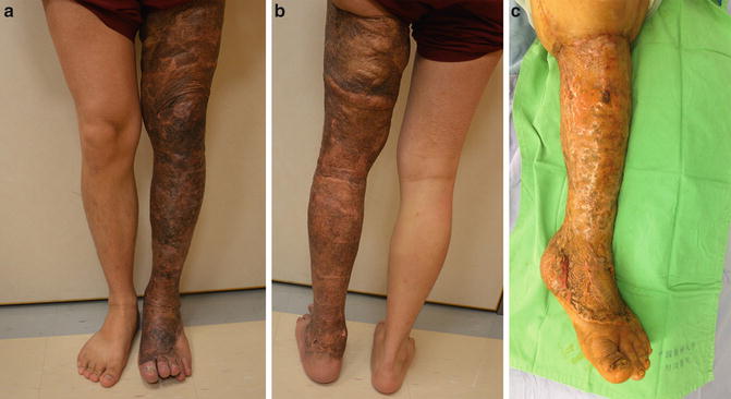
Fig. 26.2
Poor aesthetic result due to hyperpigmentation following the conventional Charles’ procedure but with good quality of the skin. (a) Anterior view; (b) Posterior view; (c) Hypertrophic scaring with fibrosis and history of ulcerations
Hung-Chi Chen (HCC)-modified Charles’ procedure combined the excisional procedure with lymph node flap transfer.
Charles’ Procedure: Surgical Technique
Preoperative Treatment
One week prior to surgery the patient is encouraged to optimize foot hygiene, have regular baths and bed rest with leg elevation. On hospital admission, a plastic surgery specialist nurse records the circumference of the affected limb. Measurements are taken at four levels: midfoot, ankle (between medial and lateral malleoli), mid-calf (15 cm below knee joint), and mid-thigh (15 cm above knee joint). Routine preoperative investigations are performed and anesthetic consultation undertaken. Antibiotics (usually cephalosporin) are administered 1 h before surgery and continued postoperatively for 3 days.
Operative Technique
Only one limb is operated at a time and the procedure is performed in one stage. The patient is positioned supine and incisions are marked. Proximally at the thigh, a circumferential incision is marked with two wedge incisions at the proximal thigh, one at the medial aspect and the other at the lateral. Excision can be limited to below knee if the thigh is relatively unaffected or on the patient’s request. Distally, at the foot and toes, markings are placed at the mid-lateral and medial aspect of the foot, above the heel and at the dorsum of the toes and web spaces, preserving the clefts between the toes. The leg will be circumferentially denuded down to fascia. Split thickness skin grafts are harvested from the entire circumference of the affected limb in a proximal to distal (axial) direction with an air-powered dermatome set at 12/1,000 of an inch. It is imperative that the lengths of the harvested STSG are as long as possible. The STSG can thus be “wrapped” around the limb in a circular fashion, so that the ends meet on the lateral aspect rather than inner aspect of the limb. Consequently, the risk of hypertrophic scar formation on the inner aspect of the limb is minimized. Furthermore, using long strips reduces the number of seams between STSG and risk of excessive hypertrophic scarring. Using long strips of STSG also avoids a patchwork appearance. The harvested STSGs can be fenestrated (but not meshed) for drainage of hematoma if necessary. A pneumatic tourniquet is placed on the proximal thigh and inflated to 375 mmHg following exsanguination. An ordinary pneumatic tourniquet cannot be used when the thigh is so large. Then a rubber tourniquet can be applied at the proximal thigh after exsanguination.
To access the posterior surface of the limb, a 3 mm pin is drilled through the distal tibia and the limb is suspended from a stand. This pin can also help elevate the leg postoperatively and is usually removed 5 days postoperatively at the bedside, before the patient is discharged.
The fibrosclerotic lymphedematous tissue is then separated from the deep fascia using blunt and sharp dissection, removing all of the soft tissue superficial to the deep fascia. The thickened deep fascia is also trimmed to its normal size.
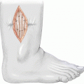
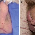
Toes are amputated if there are recurrent infections, verrucous hyperkeratosis, or osteomyelitis, as indicated in HCC-Stage IVB. Otherwise, nails and nail beds are removed, and the defect is closed using C-V/rotation-advancement flaps, to preserve length. The tip of the distal phalanx of the toe can be shortened by 0.5 cm if there is any tension during wound closure.
Once the leg has been denuded, adrenaline soaked gauze and compressive bandaging is applied and the tourniquet is deflated. All tissue excision and control of the major bleeders should be completed within 2 h of tourniquet ischemia time. Therefore, a team of surgeons is necessary.
Whilst waiting for the adrenaline to act, the proximal thigh can be debulked. Two wedge excisions are made starting at the medial and lateral mid-axial lines. The skin is undermined circumferentially and proximally to about 10 cm. The resultant flaps at anterior and posterior thigh are thinned tangentially to 2 cm and sutured together to the deep fascia to allow a smoother transition from the distal grafted thigh to the proximal thigh. Suction drains are left in situ. A full-thickness skin graft is taken from the wedge-excised tissues and used for grafting of the dorsum of the foot. FTSG on the dorsum of the foot results in more resistant skin with less hypertrophic scarring and verrucous hyperkeratosis.
The adrenaline-soaked compressive bandages are then sequentially removed in a proximal to distal direction and meticulous haemostasis is performed. The tourniquet is reinflated and the split thickness skin graft is applied circumferentially with 1 cm edge overlap. This is to prevent gap formation between the STSGs that may arise because of swelling, minimizing hypertrophic scarring. The skin grafts are applied around the ankles and knees in such a way that the seams lie on the lateral aspect of the joints.
Finally, non-adherent dressings, bulky gauze, ACE wrap, and a posterior plaster is applied. The tourniquet is then deflated. Elevation of the limb and thus avoidance of shearing force ensures STSG take on the posterior surface of the leg and thigh (Fig. 26.2).
Postoperative Treatment
During inpatient stay (5–7 days) the patient is strongly encouraged to keep the leg elevated. Deep vein thrombosis prophylaxis is administered postoperatively for Caucasian patients. Due to the low incidence of deep vein thrombosis in the Chinese population, prophylaxis of deep vein thrombosis is not needed for them [14]. Leg elevation is encouraged for the next 2 weeks and compression stocking is worn after 2 weeks. Physiotherapy and long-term limb compression are commenced subsequently (Fig. 26.3).
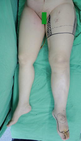
Fig. 26.3
Markings
Preservation or Amputation of Toes in Advanced Lymphedema
In advanced lymphedema, the most important goal of treatment is control or eradication of infection. The repeated episodes of infection and cellulites lead to chronic inflammation and fibrosis, resulting in occlusion of the lymphatic channels, which therefore are not amenable to any microsurgical anastomosis [3].
Toes are the major source of infection, especially in older patients [15]. Toe crowding, nail infections, skin changes such as verrucous hyperkeratosis and poor hygiene can all contribute to infection of the toes, which may ascend to involve the foot or even the proximal leg. If the toes are affected but left untreated, the patient will invariably have recurrent infections and a compromised result. Control of infection is therefore paramount. For preservation of the toes different conditions must be met in advanced stage lymphedema (HCC-Stage IVA).
(a)
The toes are swollen, but there is no history of recurrent episodes of cellulitis or appearance of verrucous hyperkeratosis.
(b)
The toes are not deformed.
(c)
There is no evidence of osteomyelitis of the toes.
The surgical technique to treat the toes includes:
(a)
Excision of the soft tissue at the dorsum of the toes with preservation of the extensor tendon and its paratenon, which can support a skin graft take. After surgery, the skin graft at the dorsum of the toes should be compressed to achieve a flat and smooth surface.
(b)
The skin flaps in the web spaces should be preserved to avoid contracture at the web spaces with resultant crowding of the toes. This can improve foot hygiene and prevent infection.
(c)
Removal of the toe nails and closure using C-V rotation advancement flaps in order to preserve length.
(d)
Amputation of toes when there is recurrent infection of the toes, verrucous hyperkeratosis, or osteomyelitis.
If the toes are affected but left untreated, the patient will have recurrent infection after performing Charles’ procedure. A recent prospective study showed that only 20 % of patients who underwent Charles’ procedure with toe amputation suffered from recurrent infections, compared to 83 % of patients who had Charles’ procedure without toes amputation. Moreover, 88 % of the latter group eventually needed toe amputation [3, 16].
Our management algorithm for treating the toes in patients with advanced lymphedema includes:
(a)
removal of nail beds (HCC-Stage IVA—with infection of nail)
(b)
removal of second and fourth toes (HCC-Stage IVB—with infection of web spaces, who want to preserve some toes)
(c)
removal of all toes (HCC-Stage IVB—with infection, who do not want to preserve toes)
Related posts:

Stay updated, free articles. Join our Telegram channel

Full access? Get Clinical Tree


