Number of patients
Mean age
Follow-up
44 pts
(41 pts w/modified technique)
1.2 years
(1 months – 10 years)
139 months
(6 months – 26 years)
The technique consists of the following steps:
1.
Opening of the abdominal wall
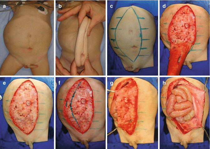
With the patient in supine position under general anesthesia, appraisal of the redundancy and flaccidity of the mid-abdominal wall is grossly calculated by the approximation of its lateral segments to the midline (Fig. 22.1). A longitudinal xipho-pubic fusiform figure is drawn on the anterior abdomen of the patient. The drawings are individually made according to abdominal flaccidity and redundancy, which vary from patient to patient and side to side. As the abdominal wall flaccidity is usually asymmetrical, the excised skin area can be more convex (lopsided) to the most flaccid side. After demarcation, the skin and subcutaneous tissue are excised, leaving the thin musculoaponeurotic fascia (MAF) exposed. The umbilicus is circumcised and left intact, attached to this fascia (Fig. 22.1). A lateral elliptical single xipho-pubic incision of the MAF is made in the most lax side, with the umbilicus being kept intact in the wider flap. This incision allows adequate abdominal access and creates one wide flap, which contains the umbilicus and one narrow flap. Individualized urinary tract reconstruction (UTR) with dissection of previous vesicostomies, nephroureterectomy, ureteral reimplantation with tailoring or infolding, cystoplasty with urachal resection and appendicular Mitrofanoff procedure to the umbilicus, and bilateral orchidopexy can be performed with excellent visceral exposure through this incision.


Fig. 22.1
(a) Initial aspect of the abdomen. (b) Appraisal of redundant abdominal wall. (c) Demarcation of fusiform figure. (d) Excising the redundant skin and subcutaneous tissue. (e) Intact MAF with increased right-sided flaccidity. (f) Single elliptical line drawn in the right side of the MAF. (g) Incision made over demarcated line providing two different sized flaps, the widest containing the preserved umbilicus. (h) Exposure of visceral contents
2.
Closure of the abdominal wall
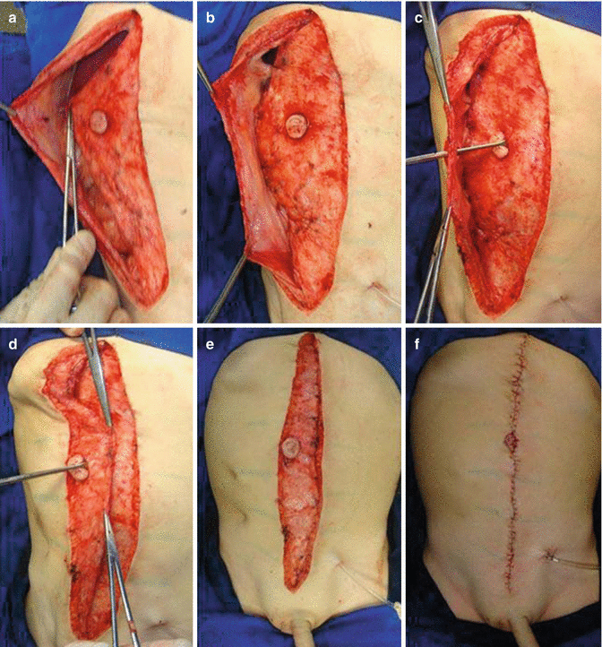
After UTR and orchidopexy, lateral fixation of the wide MAF flap to the inner side of the contralateral flap is made, therefore creating a double-layered reinforcement of the anterior abdominal wall with an outer and an inner flap, reducing its circumference and improving its tension (Fig. 22.2). Contralateral suture fixation of the outer MAF flap is then made over the inner layer. A buttonhole is made in the outer flap, in order to expose and suture-fixate the umbilicus (Fig. 22.2). Eventually, the buttonhole can be made more caudally, therefore positioning the umbilicus closer to the line that passes through the iliac crests. The skin edges are approximated medially after partial undermining of both sides. If needed, temporary urinary drainage catheters can be exteriorized through the reconstructed abdominal wall (Fig. 22.2). Likewise, a Mitrofanoff tube can be exteriorized in the umbilicus. Approximation of the subcutaneous layer and suture of the skin edges, incorporating the umbilicus, completes the procedure. Contrary to the Monfort procedure, the need to trim the skin in the upper and lower extremities of the incision is rare.


Fig. 22.2
(a) After UTR and orchidopexy, suture fixation of the wide flap to the inner side of the contralateral flap. (b) As result, creation of an inner and an outer flap. (c) Exposure of the umbilicus through the buttonhole incision of the outer flap. (d) Suture fixation of the outer flap to the inner flap (double-breasted suture). (e) Suture of the umbilicus to the orifice of the outer flap. (f) Early postoperative reconstructed abdomen, showing temporary catheter diversion
22.3 Results
Surgical complications are represented in Table 22.2. Of note, three patients presented partial ischemia of the umbilicus, which was managed conservatively without impairment of the late result. Mitrofanoff umbilical stomas were performed in three cases and remained healthy in the follow-up period.
Table 22.2
Surgical complications
Complications | Number of patients | Treatment |
|---|---|---|
Partial umbilical ischemia | 3 pts | Conservative |
Surgical infection | Ø | – |
Dehiscence | Ø | – |
Prolonged ileus | Ø | – |
Stay updated, free articles. Join our Telegram channel

Full access? Get Clinical Tree


