The understanding of the bilateral cleft lip and associated nasal deformity has evolved over the last 30 years to a point where there now exists general agreement regarding the goals, principles, and strategies for operative repair. This article presents modern tenets for repair of bilateral cleft lip and describes a logical approach to correction of the different possible subtypes.
Key points
- •
The functional and aesthetic outcome of bilateral cleft lip should be comparable with (or even surpass) that of repaired unilateral cleft lip.
- •
Modern tenets of repair include maintaining symmetry; recessing the projecting premaxilla; making accommodations for “fourth-dimensional” changes that occur with growth; constructing a full median tubercle using lateral labial elements; deepening the gingivolabial sulcus using premaxillary mucosa; establishing muscular continuity primarily; and addressing the nasal deformity synchronously.
- •
Different forms of bilateral cleft lip (eg, symmetric complete, incomplete, and lesser-form; and asymmetric) require different approaches to maintain symmetry.
- •
Although revisions of the labial repair per se should rarely be needed, small nasolabial adjustments are frequently warranted because of the secondary/tertiary stigmata that develop with growth.
Too many infants born with bilateral cleft lip undergo old-fashioned, often multi-staged, procedures, and later have to endure sundry revisions throughout childhood and adolescence. Despite the surgeon’s efforts, the stigmata of the repaired cleft lip and nose remain painfully obvious – even at a distance. To the contrary, … the appearance of a child with repaired bilateral cleft lip should be comparable to, and in many instances surpass, that of a repaired unilateral cleft lip.
Introduction
Bilateral cleft lip (bCL) has been described as the greatest of surgical challenges, and the undertaking of its correction as the most onerous of responsibilities. The rich and varied surgical history related to its repair not only illustrates the considerable difficulty of the clinical problem but also stands as a testament to plastic surgery’s ingenuity, perseverance, and indomitable spirit in its pursuit for excellence. Early, radical procedures, such as excision of the premaxilla, gave way to premaxillary setback by vomerine ostectomy and ultimately to preoperative repositioning by dentofacial orthopedics. With better understanding of blood supply and tension-reducing maneuvers, staged labial repair yielded to synchronous bilateral repair. Intricate geometric rearrangements (eg, triangular and quadrangular flaps) gave way to simpler straight line (Veau III-type) techniques, and Millard applied rotation-advancement principles. Staged procedures were accomplished safely in single stages. With deeper appreciation for growth characteristics, intentional lengthening of the prolabium (eg, König, Barsky, and others) was discontinued in favor of carefully designed, narrow philtral flaps. The relative inattention initially given to the cleft nasal deformity, other than columellar lengthening, has gradually given way to primary nasal correction and even to preoperative manipulation (ie, by nasoalveolar molding). It is by reading these stories that many important insights can be gleaned. For the so-inclined, excellent histories have been recounted by Millard and Mulliken.
Over time, there emerged a relative consensus regarding the goals, design principles, and operative strategies concerning bCL and nasal deformity. This article summarizes the contemporary approach. The article focuses on primary repair and related revisions. Palatal repair, alveolar bone grafting, and other aspects that complete the spectrum of care for these patients are presented elsewhere in this issue.
Contemporary principles
As operative strategies and techniques for repair of bCL have evolved over the last 30 years, various precepts have been clarified. These can be distilled into the following tenets for repair of the bCL:
- 1.
Maintain (or establish) symmetry
- 2.
Prepare the projecting premaxilla
- 3.
Anticipate fourth-dimensional changes that occur with growth
- 4.
Construct a full central lip using lateral labial elements and discard prolabial vermilion
- 5.
Deepen the gingivolabial sulcus using premaxillary mucosa
- 6.
Establish muscular continuity primarily
- 7.
Address the nasal deformity synchronously
Maintain (or Establish) Symmetry
Mulliken has famously written that symmetry is the one advantage that bCL has over its unilateral counterpart. Accordingly, symmetry takes pride of place as the quintessential guiding principle.
Staged repair of each side was once advocated to reduce tension on the lip, but it necessarily destroys the symmetry. Consequently, this practice should be abandoned in favor of synchronous bilateral repair. The exception to this rule is the asymmetric bCL, for which various strategies exist to improve symmetry first before undertaking labial repair. For example, in the case of a complete with incomplete CL, a nasolabial adhesion should first be performed on the greater (complete) side (in essence converting it to a symmetric bilateral incomplete CL) before attempting definitive cheiloplasty in a secondary stage.
Symmetry must be maintained throughout all stages of operative treatment; even small imperfections may become more noticeable with later growth, requiring further revisions.
Prepare the Projecting Premaxilla
The projecting premaxilla makes tension-free lip closure difficult and makes establishment of muscular continuity impossible. Recession of the premaxilla was advocated by Cronin and Monroe, among others. History credits Desault in the late eighteenth century with preoperative preparation of the premaxilla by way of linen bandages that applied external pressure over time. This might be considered the precedent for the modern process of dentofacial orthopedics, of which there are two principal methods: passive processes, such as nasoalveolar molding ; and use of active devices, such as pin-retained (Georgiade-Latham) appliances. The merits and demerits of passive and active dentofacial orthopedics are controversial and lie beyond the scope of this article, but they are considered elsewhere in this issue (see the article on nasoalveolar molding in this issue). Regardless of the method used, preoperative repositioning of the premaxilla is critical to establishing symmetry, narrowing the alveolar gap, and aligning the alveolar arches, and thus is quintessential to permitting tension-free closure of the lip, completion of the gingivoperiosteoplasty, and closure of the primary palate.
Anticipate Fourth-Dimensional Changes that Occur with Growth
Straight-line labial repairs (eg, Veau III, Manchester ) preserved all or most of the prolabial skin, resulting in an excessively wide central segment. Millard later reduced the width to better simulate a “natural” philtral column, preserving the remainder of the prolabial skin as forked flaps. However, with growth over time, the result was still a wide, shield-shaped philtrum that is one of the major secondary nasolabial stigmata.
Recent advances in the study of facial growth have not only explained this occurrence but also helped develop strategies to compensate for these changes. Using direct anthropometry, Farkas and coworkers studied craniofacial growth in developmentally normal white children aged 1 to 18 years (albeit in cross-sectional, not longitudinal, fashion). His findings reveal that some facial features are fast-growing, attaining near-adult size by age 5 years, whereas others are slow-growing. Nearly all nasolabial landmarks are fast-growing, with the notable exception of two slow-growing features: columellar length and nasal tip projection. However, philtral width (and lip in general) is a fast-growing structure. If the philtral flap is designed with normal dimensions, by age 5 years it will have widened 2.5-fold superiorly (crista philtri superioris-crista philtri superioris) and two-fold inferiorly (crista philtri inferioris-crista philtri inferioris).
Consequently, the surgeon should make accommodations for these relative growth characteristics by undercorrecting fast-growing structures and overcorrecting slow-growing structures. Mulliken discusses this at length and says that CL and nasal deformity should be corrected “in three-dimensions based on knowledge of anticipated changes in the fourth-dimension.” Also, see the article on nasoalveolar molding and the article on cleft nasal deformity and its treatment elsewhere in this issue.
Construct a Full Central Lip Using Lateral Labial Elements and Discard Prolabial Vermilion
One notable exception to the preceding rule is the median tubercle. Although it is a fast-growing structure in the normal child, it becomes slow-growing in the operated lip. Therefore, the vermilion and median tubercle specifically should be treated as a slow-growing structure and overcorrected.
Because the prolabial vermilion is thin and insufficient, it should be discarded; techniques that preserve it (eg, Manchester) should not be used because they create a “whistle deformity,” one of the more striking secondary stigmata of bCL. Rather, the central lip should be constructed from lateral labial elements and made to be as full as possible. To facilitate tension-free closure of the lateral labial elements in the midline, the soft tissue overlying the maxilla should be undermined widely in the supraperiosteal plane.
Deepen the Gingivolabial Sulcus Using Premaxillary Mucosa
Early attempts at bCL repair adapted the techniques of unilateral repair. As such, the lateral labial elements were brought toward the midline to join a prolabium/premaxilla that was largely undisturbed. A consequence of this was that the shallow central gingivolabial sulcus was left uncorrected.
Millard concluded that “methods that accept the prolabium’s attachment to premaxilla, of course, never achieve a sulcus.” As later operative strategies focused on joining the lateral labial elements in the midline behind an elevated prolabial/philtral flap, the labial mucosa creates a new anterior wall for a deepened central sulcus. To create the posterior wall, premaxillary mucosa is redraped over the premaxilla and sutured high to the periosteum. The original description of this technique is credited by Millard to DeHaan, although similar methods are also described by Mulliken and Noordhoff. An excellent history and explanation of the preferred technique was written by Marcus and coworkers.
Establish Muscular Continuity Primarily
Early attempts at establishing continuity of the orbicularis oris muscle were hampered by the projecting premaxilla and could only be accomplished in secondary stages. Modern presurgical premaxillary repositioning simplifies this challenge tremendously. Muscular continuity is essential to function (eg, puckering, whistling) and has significant aesthetic implications.
Address the Nasal Deformity Synchronously
The nasal deformity in bCL is obvious and consists of a broad, flattened nose with widened nostrils, splayed alae, recurvatum of the lower lateral cartilages (LLC), and a short columella. Early efforts either deferred nasal correction entirely or only focused on lengthening the columella by recruiting prolabial tissue (eg, forked flaps), often in delayed or staged fashion. Eventually, the old perception that “prolabial skin belongs to the columella” was replaced by a new mantra, “the columella is in the nose” ; that is, by directly addressing aspects of the nasal deformity per se, the columella can be lengthened. Early proponents of primary correction of the CL nasal deformity include Broadbent and Woolf, McComb, Mulliken, Trott and Mohan, and Cutting and coworkers. Both open and semiopen (“closed”) approaches have been described, and the use of preoperative nasoalveolar molding. Pigott assessed nasolabial aesthetic outcome of primary nasal correction versus no nasal correction (in unilateral CL) using a panel-based evaluation method, showing significantly improved symmetry and aesthetic outcome with primary repair.
Repair of bilateral complete cleft lip
Contemporary technique for repair of bilateral complete CL in the style of Mulliken is presented next. A superb and expertly illustrated explanation of all steps is available. Alternative approaches to bCL repair have also been described by Chen and Noordhoff, Byrd and coworkers, and Monson and coworkers.
Markings
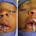
A double-ball retractor placed in the nostrils to gently elevate the nasal tip is useful to preserve symmetry while facilitating visualization. Marking begins with identification of standard anthropometric reference points, including nasion (n), pronasale (prn), columella nasi (c), subnasale (sn), ala nasi (al), subalaris (sbal), labiale superius (ls), crista philtri superioris (cphs), crista philtri inferioris (cphi), stomion (sto), chelion (ch), and endocanthion (en). When taking measurements for symmetry, point sn serves as the main reference for the prolabial markings, and point sbal serves as the main reference for the lateral lip markings. The junction between vermilion and labial mucosa (“red line”) should also be indicated with a dotted line ( Fig. 1 ).
On the prolabium, the philtral flap is drawn. For a child younger than 1 year of age, the philtral flap should measure 6 to 7 mm in vertical height (sn-ls), which is often the total available height of the cutaneous prolabium. The philtral flap should be made 2-mm wide at its superior aspect (cphs-cphs) and 4-mm wide at its inferior aspect (cphi-cphi), and its sides should bow slightly inward. Extracutaneous prolabium on each side of the philtral flap may be de-epithelialized to create “flanking flaps” measuring approximately 2 to 3 mm in width. These flanking flaps provide the extra bulk needed to simulate a philtral ridge when buried under the advanced lateral labial flaps (for clarity in the markings, the flanking flaps are usually denoted with a striped pattern). The remainder of the prolabial skin and vermilion are discarded. Premaxillary mucosa is used to construct the posterior lining of the gingivolabial sulcus.
On each lateral lip element, the peak of Cupid’s bow (cphi′) is placed as far medial as possible where there is still adequate vermilion for the median tubercle and where at least 3 mm of well-defined white roll still exists medially. It is important not to choose a point too medially, where the white roll has already become indistinct or where the vermilion has begun to taper; doing so has been shown to correlate with retained cleft tissue within the repair and poorer aesthetic and functional outcomes. The lateral labial advancement flap on each side is drawn as large as possible, noting that it is trimmed later during inset and closure. It is marked medially following the mucosal-cutaneous junction and inferiorly along the vermilion-cutaneous border, just above the white roll, extending to the marked cphi′ point. Superiorly, it curves around the alar base and extends up to, or just beyond, point sbal. It is important that this mark stay just above the crease of the normal alar base-labial junction.
A dilute solution of lidocaine and bupivacaine with epinephrine is infiltrated. Key landmarks (eg, chpi′) are tattooed, and a scalpel may be used to score the markings.
Labial Dissection
A mini-blade or #15c scalpel is used to incise the markings of the philtral flap, and the flanking flaps are de-epithelialized. The remaining prolabial skin is discarded. Using a fine double hook and sharp scissors, the philtral flap is elevated (en bloc with the paired flanking flaps) in the subcutaneous plane to the level of the anterior nasal spine.
The lateral labial flaps are incised. Sharp scissors are used to separate the vermilion-mucosal flaps from the musculocutaneous portion. After this is accomplished, sharp dissection is continued beneath the muscle down to the maxilla, and a Tessier elevator is used to widely mobilize the soft tissue in the supraperiosteal plane as far as the malar eminences. Wide mobilization is a critical maneuver to reduce tension on the midline mucosal, muscle, and vermilion closure. Next, the scalpel or sharp scissors are used to dissect free the orbicularis oris muscle from overlying skin and underlying mucosa for a distance of approximately 1 cm.
Closure of the Nasal Floor
Medial and lateral nasal mucosal flaps are elevated from the premaxilla and inferior turbinates, respectively, and closure of the nasal floor is begun. Later in the procedure, the alar base flaps are transposed medially and sutured to the C-flaps, completing the closure of the nasal floor.
Gingivoperiosteoplasty and Mucosal Flap for Deepening of the Gingivolabial Sulcus
Previously, the philtral flap and paired flanking flaps were elevated in the subcutaneous plane, and excess prolabial skin was excised. At this point, the thin strip of prolabial vermilion is removed, and the premaxillary mucosa is cleaned of any residual subcutaneous fat. The gingiva and periosteum of the lateral alveolar segments are then incised vertically, and gingivoperiosteoplasty is performed using chromic suture, beginning posteriorly and proceeding anteriorly. Gentle pressure on the premaxilla may be applied to facilitate closure. Subsequently, the remaining inferiorly based mucosal flap is pulled upward and secured to the premaxillary periosteum using polydioxanone (PDS) suture. This forms the new posterior wall of a deepened gingivolabial sulcus ( Fig. 2 ).
Related posts:


Stay updated, free articles. Join our Telegram channel

Full access? Get Clinical Tree


