Microvascular free tissue transfer is the best modality of replacing composite tissue defects with composite vascularized tissue. Wound healing, functional reconstruction, rehabilitation, and cosmesis are best accomplished when the tissue defect is replaced by free tissue. The reconstructive tissue can be tailored to the defect and is harvested from outside the often radiated pretreated reconstructive field. Evidence to support the use of free tissue transfer in head and neck defects is not of the highest level. This article reviews the postoperative monitoring of free tissue transfer, lateral mandibular reconstruction (fibula vs radial forearm), and functional outcomes with free tissue transfers.
Key points
- •
The goal of evidence-based medicine is to evaluate outcomes to provide improved care to patients.
- •
In the field of microvascular reconstruction, the level of evidence is mediocre.
- •
Monitoring of free flaps has allowed for improved outcomes and lower revision rates.
- •
Reconstruction with bony flaps for the lateral mandibular defect has proven to be efficacious.
- •
A collaborative, multiinstitutional study should be able to improve the level of evidence that exists.
Introduction
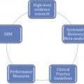
Evidence-based medicine integrates research, clinical expertise, and patient values. It attempts to evaluate scientific evidence and clinical care experiences to provide a rationale for decision making in the care of patients. This contrasts with the tradition of treating patients as was learned in residency or fellowship. It strives to bring a rationale to the decision-making process as opposed to the commonly quoted, “In my experience.” Finding good literature to make evidence-based decisions when it comes to operative procedures is difficult. Randomized, controlled studies, considered the best modality to amass evidence, are almost impossible to perform when comparing 2 different operative procedures, or across modalities of treatment focused on low volume-disease, such as head and neck cancer. For a variety of reasons, we are often left with comparing different procedures as they have evolved in a single institution or with comparing results between institutions. Still, it is possible to use this broad-based information if we recognize the selection biases and the limitations of the data.
In this article, we present 3 areas in the field of microvascular reconstruction where data exist that allows one to make an evidence-based decision on clinical care. The first is postoperative monitoring of free tissue transfer. Although it seems logical and empiric that monitoring is a good thing to do, little clear-cut evidence exists as to its efficacy. Second, the reconstruction of lateral mandibular defects can be performed in a number of ways. Few comparative studies have been performed, yet some evidence does exist. Finally, functional outcomes are reported to be improved with free tissue transfer. Analysis of the individual surgeon or institutional evidence reveals superiority of free tissue over regional or local transfer, yet functional outcome reporting is inconsistent with most reports using nonvalidated instruments or custom designed assessment tools. Thus, this review sought to define the state of reporting for functional outcomes in head and neck reconstruction studies suggesting they provide function information, and assess quality of life (QOL) data between 2002 and 2012.
Introduction
Evidence-based medicine integrates research, clinical expertise, and patient values. It attempts to evaluate scientific evidence and clinical care experiences to provide a rationale for decision making in the care of patients. This contrasts with the tradition of treating patients as was learned in residency or fellowship. It strives to bring a rationale to the decision-making process as opposed to the commonly quoted, “In my experience.” Finding good literature to make evidence-based decisions when it comes to operative procedures is difficult. Randomized, controlled studies, considered the best modality to amass evidence, are almost impossible to perform when comparing 2 different operative procedures, or across modalities of treatment focused on low volume-disease, such as head and neck cancer. For a variety of reasons, we are often left with comparing different procedures as they have evolved in a single institution or with comparing results between institutions. Still, it is possible to use this broad-based information if we recognize the selection biases and the limitations of the data.
In this article, we present 3 areas in the field of microvascular reconstruction where data exist that allows one to make an evidence-based decision on clinical care. The first is postoperative monitoring of free tissue transfer. Although it seems logical and empiric that monitoring is a good thing to do, little clear-cut evidence exists as to its efficacy. Second, the reconstruction of lateral mandibular defects can be performed in a number of ways. Few comparative studies have been performed, yet some evidence does exist. Finally, functional outcomes are reported to be improved with free tissue transfer. Analysis of the individual surgeon or institutional evidence reveals superiority of free tissue over regional or local transfer, yet functional outcome reporting is inconsistent with most reports using nonvalidated instruments or custom designed assessment tools. Thus, this review sought to define the state of reporting for functional outcomes in head and neck reconstruction studies suggesting they provide function information, and assess quality of life (QOL) data between 2002 and 2012.
Monitoring free tissue transfer
The ability to transfer successfully tissue from 1 part of the body to another is related to patient factors and the technical ability of the microvascular reconstructive surgeon. Total failure of the flap is a rare and devastating complication that is usually secondary to vascular anastomotic failure. This results in thrombosis of the anastomosis and death of the tissue. The need to reexplore microvascular anastomosis is consistently reported at between 10% and 12% (level 4 evidence). When flaps are explored in a timely manner, successful revascularization is reported it up to 70% to 80% of cases. This led to a belief that monitoring of the tissue in the postoperative period should be intensive. Routines have involved clinical monitoring the flap hourly by medical personnel. The advent of resident duty-hour restrictions and improved technology has changed the paradigm so that nursing personnel primarily monitors the flap with medical input on a less frequent basis.
Clinical Monitoring Involving Evaluation of the Flap by Physical Examination
Looking for color, turgor, and warmth has been the mainstay of physical examination for many decades ( Fig. 1 ). Even with this modality, the incidence of flap revision and ultimately survival has not changed (level 4 evidence). This led to the desire to find technological methodologies to supplement the clinical examination. By far the most common method used is the placement of an intraoperative Doppler monitor (level 4 evidence).
The Doppler can be placed in a sheath around the artery or it can be used as part of an implantable coupling device ( Fig. 2 ). Schmulder and colleagues in 2011 compared 259 patients monitored with the implantable Doppler and compared them with 289 patients monitored using clinical measures only (Level 3 Evidence). The patients monitored with the implantable Doppler had an overall survival rate that was significantly higher than those monitored clinically. The reexploration rate was also higher in the flaps with implantable Dopplers. The surgical salvage rate improved from 40% to 95% and the success rate improve from 84% to 95%. False positives occurred in 3 of 36 patients. In 2 of these, the patient was explored and the Doppler wire was dislodged. There were no false negatives. Most authors suggest that, when the Doppler suggests compromise and the clinical examination suggests a viable flap, careful and intensive observation can be performed after trouble shooting the device.
Wax evaluated 1142 patients who had a Doppler placed in the intraoperative setting. They determined that 10% of patients develop intraoperative vascular problems that were successfully revised. Compared with contemporary literature with a reexploration rate of 10% to 14%, their postoperative revision rate was 7%. The majority of revisions were after 12 hours, in contradiction to the literature in which the majority of revisions are within the first 12 hours. They felt that use of the Doppler in the intraoperative setting allowed them to detect issues with the vascular anastomosis that would have been undetected until the patient was in the recovery room or on the ward. They also demonstrated that the patients who had intraoperative vascular issues were more likely to suffer postoperative issues with a lower salvage rate.
Overall, this seems to indicate that monitoring of free flaps for vascular compromise in the intraoperative and postoperative setting is beneficial to patient care. The detection of vascular compromise allows for an active intervention with salvage in a significant number of patients (level 4 evidence).
Lateral mandibular defect
Reconstruction of the mandible continues to be a challenging problem faced by reconstructive surgeons. Techniques have evolved over the last century. The mandibular swing was the standard operation with adequate, although improvable, healing and functional outcomes. The cosmetic outcomes were poor and dental rehabilitation was not possible ( Fig. 3 ). The introduction of rigid fixation allowed surgeons to improve rehabilitation. With improved materials, a lateral mandibular defect could be reconstructed safely with local tissue transfer and a plate. Although this had proved to be successful in many cases, over time extrusion or fracture of the plate would happen (level 4 evidence). The patients were also unable to undergo full dental rehabilitation and plate extrusion rates were higher than was desirable. Reconstruction continued to evolve and free tissue transfer allowed for the replacement of the composite bony defect, allowing for oral and dental rehabilitation. It is now purported that bony reconstruction is the “gold” standard (level 3 evidence).
The choice of which osteocutaneous free tissue transfer to use has been complex. For lateral mandibular defects, the radial forearm osteocutaneous ( Fig. 4 ), the fibula osteocutaneous ( Fig. 5 ), and scapula osteocutaneous ( Fig. 6 ) are most commonly used (level 3 and level 4 evidence). The fibula osteocutaneous free flap his evolved as the best reconstructive modality owing to its ability to handle dental implants and thus improve dental rehabilitation. Although this is stated empirically, it has been difficult to prove. The study by Dean and colleagues (level 3 evidence) compared outcomes of patients reconstructed with radial forearm osteocutaneous flaps as opposed to the fibula osteocutaneous flap. They determined that the outcome was equivalent between both groups in terms of postoperative diet, return of oral function, and oral rehabilitation. Others have substantiated this result. Interestingly, none of the fibula patients obtained implants for dental rehabilitation and only a few obtained dentures. This demonstrated the disconnect seen between theoretic benefits as speculated on by the surgeon and what patients are willing to follow through on. Although mandibular reconstruction with free tissue transfer is the best option for healing, oral rehabilitation, and cosmesis in this patient population, the goal of dental rehabilitation remains elusive. Whether a fibula or radial osteocutaneous flap provides the best reconstruction is unknown at this time. If dental implants are to be used, then the fibula is the best option; however, the data are lacking.
Functional Outcomes in Reconstruction of the Oral Cavity and the Oropharynx
The impact of microvascular free tissue transfer in reconstructing ablative defects of the oral cavity and the oropharynx has significantly shaped the surgical management of malignancies in these subsites. With the proven reliability of free tissue transfer, the future emphasis will be on analyzing and optimizing function. Evaluations of functional outcomes are reported in several ways: (1) examination of a particular flap/technique in reconstructing defects, or (2) by evaluating a surgeon’s experience with a flap or comparison of different flaps in reconstruction of similar defects. Within these studies, there is a diversity of metrics ranging from aesthetic-related questions and functional objective data to QOL questionnaires. Furthermore, the consistency of metrics is variable, ranging from self-created outcome measures to validated instruments. In general, there is a lack of clear consensus on which measures to use; as a result, comparisons between studies is challenging.
Systematic reviews are structured literature reviews characterized by robust methodology and a focused research question (level 5 evidence). Systematic reviews in head and neck reconstruction have increased in recent years, but a definitive review of the functional outcomes in reconstruction is lacking (level 3 evidence). Although nonoperative management of head and neck cancers have well-designed, single-institution studies as well as systematic review to assess functional outcomes (level 3 evidence), such rigor in examining the role of reconstruction in the operative management of this disease has not been employed. We asked the following question to guide our systematic review: what are the functional outcomes of head and neck reconstruction involving the oral cavity and oropharynx? The primary aim of the review was to understand the functional implications involving reconstructions of the oral cavity and oropharynx; the secondary aim was to examine the metrics used in examining functional outcomes. Our objective was to assess the current state of research regarding functional outcomes in reconstruction of oral cavity and oropharyngeal defects, assess the limitations of the studies, and propose recommendations on how to guide further research on functional outcomes.
Historically, free tissue reconstruction after ablative surgery was thought to be associated with worse functional outcome than primary closure. McConnel and colleagues argued that oropharyngeal defects were best closed with a primary closure. However, close inspection of the data reveals that the defect volume/reconstruction volume was higher in local reconstruction versus free tissue; this technique would leave a deficit even in resected and reconstructed beds that do not approximate functional needs as they are now understood. Free tissue should reconstitute at least the volume of lost tissue with some overcorrection to account for shrinking and radiation change (level 3 evidence).
Comparisons between regional flap reconstructions, such as pectoralis major flaps and free tissue for oral and oropharynx sites, reveal superiority of free tissue (level 3 evidence). Hsing and colleagues compared QOL outcomes in 491 consecutive patients undergoing resection and regional transfer to free tissue reconstruction of the oral cavity. Applying the University of Wisconsin QOL survey, they were able to demonstrate that patients receiving free tissue rated their speech, mood, and shoulder function domains significantly better than those that had pectoralis rotation flaps. In addition Guerin-Lebailly and colleagues showed that patient, family, and professional assessments of speech were superior in their cohort of free tissue reconstructed patients compared with local or regional tissues; sentence intelligibility, food score, and Hirose scoring outcome (a measure of speech function) were significantly better with free flaps for oral cavity defects (level 3 evidence). Markkanen-Leppanen and colleagues in a series of studies further demonstrated that oral and oropharynx defects compromise velopalatal dynamics and alter speech articulations, but that free tissue can help to improve negative aspects. They also demonstrated that treatment negatively impacted QOL at 12 months, and this was reportedly associated with socioeconomic status. Specifically, voice quality remained the same after free tissue but articulations, particularly of /r/ and /s/ proved challenging.
Because free tissue has become the mainstay of oral and oropharynx upfront reconstructions, more literature has emerged reporting functional information. Complex interplay between tumor type, site, prior or future therapy, type of reconstruction, skill level of reconstructive surgeon, motivation of patient for rehabilitation, skill of speech therapist all affect the dynamic.
Related posts:

Stay updated, free articles. Join our Telegram channel

Full access? Get Clinical Tree


