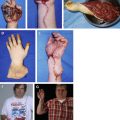
Treatment of complex hand trauma includes adequate debridement of nonviable tissue, early reconstruction, and careful selection of various available surgical procedures tailored to patients’ needs and requests. Debridement of all necrotic tissue is crucial before any attempt at reconstruction. Surgeons should also consider cosmetic outcomes of the reconstructed hand and donor-site morbidity. For best results reconstruction should be performed early, with proper early postoperative therapy. This article reviews the principles and surgical options in the management of complex hand injuries involving the dorsal and the palmar aspects of the hand, and the different types of tissue in the hand.
Key points
- •
Debridement of all necrotic tissue is crucial before any attempt at reconstruction.
- •
If vital structures are exposed, timing of reconstruction is important in planning.
- •
Preoperative planning should identify an appropriate flap for tissue coverage.
- •
Cutaneous defect coverage is as follows. Dorsum of the hand (<20 cm 2 ): regional pedicle flap (radial forearm flap; posterior interosseous flap; distal ulnar and radial artery perforator flaps). Dorsum of the hand (>20 cm 2 ): radial forearm fasciosubcutaneous flap, anterior lateral thigh flap, lateral arm flap, groin flap (pedicle or free), partial muscle flaps (latissimus dorsi or rectus). Palm of the hand: regional pedicle flap for the thenar eminence, and medial plantar flap for the hypothenar eminence and central region.
- •
For composite defect coverage, tendocutaneous defects are best managed with single-stage reconstruction (tendon grafts in conjunction with pedicle or free flaps, or with composite tendocutaneous flaps). Osteocutaneous defects may be treated with a composite flap (radial osteocutaneous flap or fibular osteoseptocutaneous flap); the choice is made strictly according to the size and location of the defect in the hand.
Introduction
The treatment of industrial and vehicular hand trauma remains a challenge for hand surgeons. Complex hand trauma often requires tissue coverage associated with tendon, bone, and joint reconstruction. The introduction of microsurgery has totally modified the surgical approach to this type of injury, allowing for early debridement with immediate, simultaneous reconstruction and soft-tissue coverage. The aim of this article is to provide guidelines for the surgical approach to complex hand trauma.
Debridement
Debridement of all necrotic tissue is crucial before any attempt at reconstruction. Many investigators recommend early aggressive debridement rather than traditional serial debridement; the concept of waiting for the development of good granulation tissue is confined to the past. As granulation tissue forms, distinguishing among tissue layers becomes less clear. Moreover, granulation tissue can become infected, increasing the risk of developing joint stiffness and tendon adhesions. These changes delay early mobilization of the hand, which is fundamental to restoring function. Serial debridement can be considered only in particular circumstances, such as electrical burn injuries or severe crush injuries, and in unstable patients and those with massive wound contamination. In these cases, initially estimating the extent of tissue damage can be difficult.
When edema appears, usually 3 to 4 days after initial injury, all tissues tend to indurate. Muscle, tendon, ligament, and joint are involved in this process, concomitant with the risk of later joint stiffness and tendon adhesions.
Radical debridement is performed under tourniquet control. The authors’ principle is to start from outside the injured area to recognize and preserve the important structures (nerves, tendons, vessels). Crushed and contaminated skin, avulsed and mangled tendon, and devascularized muscle must all be excised. The same principle has to be applied to free bony fragments without soft-tissue attachment. When all the devitalized tissues are removed, the tourniquet is released and the wound is irrigated with antibiotic solution. If necessary, the debridement is continued until every part presents satisfactory bleeding.
The following are 2 examples of injuries to dorsum of the hand treated elsewhere by waiting for granulation tissue to develop, which became more difficult to treat later. A 32-year-old man sustained a severe abrasion injury to the dorsum of the right hand and was referred 8 days after injury. The dorsum of the hand was entirely covered by granulation tissue; on radical debridement there was tendon loss in the middle, ring, and small fingers, and avulsion of the interosseous muscles in the third and fourth metacarpal spaces ( Fig. 1 ). The second case concerned a 52-year-old man with a long history of infection localized to the dorsum of the left hand. The suggested management is to remove all the dead or contaminated tissue following the approach proposed by Godina : “debridement should be done tumor-like, en bloc wound excision,” leaving behind a bed that is ready for immediate coverage ( Fig. 2 ).
Timing of reconstruction
Early closure of wounds through the use of tissue transfer was initially introduced at the end of the 1980s. A free flap is considered early if it is performed within 72 hours. If this delay is reduced to the first 24 hours, the flap is called an emergency free flap. The choice of one surgical approach over the other strictly depends on the presence of exposed vital structures. A vein graft restoring vascular supply should not be left exposed, and is an absolute indication for immediate primary coverage; exposed hardware, in addition to exposed nerve or tendon repairs, also need immediate coverage.
Use of nonreplantable parts to reconstruct significant structures is an absolute indication for emergency reconstruction. Relative indications for immediate reconstruction are exposed and, as yet, unrepaired tendons, nerves, joints, and bones. These structures can tolerate only a short period of exposure before they are at risk of permanent loss. The ideal timing for coverage of such structures should be within 72 hours of injury. However, in the authors’ practice it is not rare for reconstruction to happen after 3 to 5 days, sometimes even after 1 week. This delay is a consequence of initial treatment or emergency surgical repair taking place at outside facilities, often by inexperienced surgeons, before referral to a specialized hand center.
The following case illustrates how such injuries should be handled sequentially. A 29-year-old man reported a severe crush injury with skin avulsion involving the thumb and the fingers of the right hand ( Fig. 3 A). Owing to its bad condition, the index finger was used as a “bank finger.” The dorsal skin of the first phalanx was transferred, as an island flap (kite flap), to cover the large pulp defect of the thumb. The palmar skin of the first phalanx was used as a free flap to wrap the pulp defect of the ring finger (see Fig. 3 B). The index finger was amputated through the metacarpal bone. The final result shows an “acceptable hand” with 3 fingers, 2 of which have an almost normal length, and a fully functioning thumb, all with near-normal sensibility (see Fig. 3 C).
Preoperative planning
In complex hand trauma, the need for soft-tissue coverage should be evaluated carefully. Every patient must be assessed based on age, sex, occupation, educational level, motivation, concomitant injuries, and predicted postoperative compliance with physiotherapy. Weighing all of these criteria, it is possible to tailor a reconstructive plan to each individual patient.
Free tissue transfer is indicated when local or distant pedicle flaps from the fingers, the hand, or the forearm cannot be harvested owing to the extent of injury or the involvement of major vessels. Free flaps can be used in complex hand injuries because it is possible to transfer multiple vascularized tissues in a single-stage procedure.
The flap selected for reconstruction should match the size, shape, location, color, and texture of the tissue surrounding the defect; for example, the skin of the palmar surface of the hand is very different from dorsal or finger skin. Donor-site defects are also highly important in both female and male patients. Nowadays patients only accept the donor-site morbidity that results in limited functional disability. To avoid patient dissatisfaction, it is advisable to involve patients in preoperative decision making and to inform them about the types of treatment, the predicted disabilities, and the donor-site morbidities. Correct flap selection is the first step toward achieving good results.
It is also important to remember that one of the general principles in complex hand trauma concerns the assessment of hand perfusion. The absence of adequate blood supply is assessed by direct clinical observation; an angiogram is rarely necessary because simple Doppler ultrasonography can ensure satisfactory information about the vascular anatomy of the hand; in fact it can be used to identify vessels for possible free flap anastomoses, verify the presence of an intact palmar arch if a pedicled regional flap is planned, or identify a perforator vessel if a perforator flap is designed.
Cutaneous defect coverage
Many flaps are currently available for hand coverage. The choice is strictly based on the location and the size of the defect.
Dorsum of the Hand
The dorsum of the hand is a specialized region with thin and fragile skin characterized by poor subcutaneous tissue. The dorsal aspect of the hand is frequently prone to different types of injuries (crush, degloving, hot press, friction, and so forth) that result in exposed tendon and bone. The treatment of cutaneous defects may be achieved with local pedicle flaps, distant pedicle flaps, or free flaps. The choice of which technique to use depends mainly on the size of the defect. For medium-sized defects (<20 cm 2 ) pedicle flaps are a simple solution, offering pliable skin very similar to that of the dorsum of the hand.
The options for regional pedicle flaps include the radial forearm flap, the ulnar forearm flap, the posterior interosseous flap, and the more recently described distal ulnar and radial artery perforator-based flaps.
The radial forearm flap is suitable for coverage of the dorsal aspect of the hand; its use has been criticized because it requires the sacrifice of a major artery in an already traumatized hand, and because the donor-site appearance is not always satisfactory. It may have a role in hand reconstruction, however, if used to cover medium-sized defects with direct closure of the donor site and without jeopardizing the vascular supply to the hand.
Ulnar pedicle and posterior interosseous flaps have not gained the same degree of consensus. This opinion is probably based on concern surrounding harvesting the more dominant ulnar artery with the ulnar forearm flap. The posterior interosseous pedicle island flap has some advantages over the radial forearm flap: it is thinner, there is less morbidity at the donor site, and the major artery is preserved. Its greatest drawback is the limited sizes available for the flap, as closure of the donor site is successful only if it is less than 3 to 4 cm wide; otherwise, skin grafting the donor site often results in poor cosmetic outcome ( Fig. 4 ).
The authors consider these pedicle flaps to be still useful, particularly for young surgeons taking night call. In their practice the posterior interosseous and radial forearm flaps are used only when the donor site is able to be closed primarily without using a skin graft.
A 47-year-old man presented with a severe shotgun injury to the dorsum of the left hand with fractures of the second, third, and fourth metacarpal bones ( Fig. 5 A). The index and middle metacarpal bones needed stabilization plates and the ring metacarpal required multiple screws. After debridement, the extensor tendons were exposed and coverage was performed using a reverse pedicle radial forearm flap (see Fig. 5 B); the donor site was closed directly. The final result had a good cosmetic and functional outcome (see Fig. 5 C), as the color and texture of the flap matched that of the normal hand and there was an acceptable donor-site result. Of equal importance, this flap did not require surgical revision.
Perforator flaps, based on perforators of the radial or ulnar artery, are harvested from the forearm as either fasciocutaneous or adipofascial flaps, the latter usually requiring a simultaneous full-thickness skin graft. These flaps have some disadvantages: they can only cover moderate-sized defects and their pivot points are proximal to those of traditional pedicle flaps, making them less suitable for defects in the distal dorsal aspect of the hand. In addition, the adipofascial pedicle is relatively bulky after rotation, making direct closure a danger to the pedicle. If planning these flaps, it is critical to consider possible damage to the radial or ulnar artery perforators in cases of complex injury to the forearm.
The use of pedicle flaps is more restricted in cases characterized by large skin defects (more than 20 cm 2 ). After raising a pedicle radial forearm flap, the donor-skin defect has been criticized because of the poor donor-site result ( Fig. 6 ). Outcomes can be partially improved by using the retrograde radial forearm adipofascial flap, which removes only the fascia and fat layers of the forearm tissue, leaving forearm skin intact. Another possibility is to divide the radial forearm flap into different sections based of the perforators and using a long, narrow flap, allowing for primary closure of the donor site.
Fasciocutaneous flaps are the most commonly used flaps for the management of large defects of the dorsal hand. The choice of which flap to use depends on many factors: the cosmetic match of the skin surrounding the defect, the reduction of donor-site morbidity, and the approach of simultaneous wound debridement and flap harvesting. The anterolateral thigh flap has recently received attention from hand surgeons, and now represents a good alternative to other fasciocutaneous flaps. The lateral arm flap is sometimes limited by its short pedicle. The scapular flap requires a change of position during the operation, hindering a 2-team approach. Compared with these flaps, the anterolateral thigh flap has numerous advantages: simultaneous flap elevation and preparation, shorter operative time, longer vascular pedicle (approximately 10 cm long), and the option to harvest a large skin paddle even when basing the flap on a single perforator. Before it is transferred on the hand, this flap can be thinned to approximately 3 to 4 mm by removing a considerable amount of fatty tissue. This procedure is important in obtaining an optimal match between the donor tissue and the area to be reconstructed with the flap.
A 22-year-old man sustained a severe hot-press injury on the dorsum of the right hand, and was treated 20 days after the initial trauma. After debridement, a large anterolateral thigh flap was raised from the left thigh ( Fig. 7 A). The donor defect was covered with a split-thickness skin graft. Although the flap was initially thinned, additional debulking was performed successfully to improve the final cosmetic result (see Fig. 7 B).
Another useful technique is the groin flap, which can be either a pedicle or a free flap. The free groin flap presents some advantages: it does not require immobilization of the hand, it can be raised as a large flap, and it can be harvested very thinly.
Instead of fasciocutaneous flaps, some investigators suggest the use of muscle flaps covered with a split-thickness skin graft. Muscle flaps are tailored to the size of the defect; the most commonly used are the partial superior latissimus or medial rectus flaps. With these flaps, debulking usually is not necessary, and good cosmetic results are achieved in the hand as well as the donor site, as most of the donor muscle remains intact. The authors’ experience with muscle flaps is still confined to those cases characterized by defects with a dead space or previous severe infection.
Palm of the Hand
The palm of the hand is an important instrument in interacting with the environment. The skin is even more specialized than that of the dorsum. While covering deep structures such as tendons, vessels, and bones, it is responsible for processing sophisticated tactile information, and simultaneously must sustain the mechanical stress of everyday activity. To assure efficient prehension of the hand, the skin must be stable and able to absorb and distribute shearing stress and pressure. For this reason it is provided with special fibrous septa, which connect the derma to the palmar fascia to allow a firm grasp. In addition, the high density of the Pacinian corpuscles accounts for the unique sensibility of the palm of the hand. Tubiana introduced the concept of “functional cutaneous units” in the palm of the hand, taking into account mobility versus stability; he concluded that the hypothenar and central units are the more stable and fixed, whereas the thenar region is provided with more mobile skin. Engelhardt and colleagues analyzed the palm of the hand from the sensibility standpoint and realized that, after the pulp of the fingers and thumb, the thenar and hypothenar units are the most important regions for light touch.
Such complex anatomy and function of palmar tissue are difficult to reconstruct. Although the palm of the hand has a great potential for spontaneous repair, as demonstrated by the feasibility of the open palm technique for severe Dupuytren disease, coverage with vascularized tissue is mandatory when tendons or neurovascular structures are exposed. Fasciocutaneous pedicle flaps from the forearm were extensively used in past years to reconstruct palmar tissue. These flaps have been replaced to some degree by local perforator-based flaps, among which the dorsal ulnar perforator flap has been proved to be particularly useful. This flap comes from the Becker flap, which may be considered its precursor. To lift it, the dorsal ulnar branch of the ulnar artery is dissected under magnification to its origin at the main vessel ( Fig. 8 A). This dissection allows a significant increase in the arch of rotation, opening the possibility to cover the most distal region of the palm with very low impact to the donor site (see Fig. 8 B).