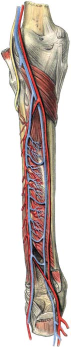
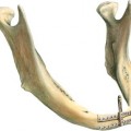
36 Microsurgical Anastomosis of the Fibula Bone Reconstruction of mandibular and maxillary defects represents a challenge to the oral and maxillofacial surgeon. The restoration of form and function is paramount for rehabilitation of the affected patient. As a consequence, the shape and size of the graft should correspond with the bone segment that has to be replaced. The vascular pedicle should be long, constant, and offer the possibility of concomitant transfer of soft tissue for replacement of mucosal and cutaneous defects. To facilitate masticatory rehabilitation by the placement of dental implants, the grafts should consist of a thick cortical plate surrounding cancellous components of high density. Vertical bone height of 7–10 mm and a bone width of 6 mm are required as a minimal volume. To date, the fibula flap is one of the most popular options for bony reconstruction in oral and maxillofacial surgery. It is the workhorse of modern-day mandibular reconstruction. Free vascularized osseous or osteocutaneous flaps can be adopted for the reconstruction of extensive bony and soft-tissue defects. The fibula allows division into segments using a wedge osteotomy technique to precisely recreate the shape of the resected bone. In contrast to the homogenous structure of the alveolus, the fibula is the best flap to be harvested in obese patients. There is only limited donor site morbidity. It offers a reliable donor site. However, variations in vascularization of the fibula are possible, and thus preoperative angiography or color Doppler ultrasonography is recommended. The fibula is composed of heterogeneous portions consisting of a dense cortical margin and a small cancellous core. The placement of implants into dense fibular free flaps results in a high primary stability. Sometimes primary stability is high enough to permit immediate functional loading of these implants (Chen, Chen, and Hahn, 1994; Hidalgo, 1989, 1991, 1994; Hidalgo and Pusic, 2002; Hidalgo and Rekow, 1995; Hausamen and Neukam, 1994; Neukam and Hausamen, 1996; Neukam, Schmelzeisen, and Schliephake, 1994; Schmelzeisen et al., 1996; Peled et al., 2005). The fibula is a straight bone that is up to 40 cm long. To maintain the integrity of the knee and ankle joints, 7 cm of bone proximally to the neck of the fibula and distally to the lateral malleous have to be preserved. Segments of fibular bone up to 26 cm in length can be harvested. Segmentalization of the fibula to restore symphyseal contour and jaw relationship with a view to future implant rehabilitation is a demanding aspect of the reconstruction because of the inherent risks of devascularization, instability, and malposition of the segments (Baehr et al., 1998). However, wedge excision osteotomy is a reliable technique to achieve the required shape of the graft. One disadvantage of the fibula graft is its limited vertical height that may cause problems during prosthetic reconstruction (Klespers et al., 2000). If placed at the caudal border of the mandible, the distance between implant shoulder and the occlusal plane is large, leading to unfavorable crown-to-implant ratios. Doubling of the fibula helps to mitigate this problem (Baehr et al., 1998). An alternative method is vertical distraction osteogenesis of the graft (Chiapasco et al., 2000). The peroneal artery provides the blood supply of the fibula by an endosteal nutrient medullary artery as well as periosteal circulation (Fig. 36.1). The graft can be raised as a free osseous or a free osteocutaneous flap with a thin pliable paddle of skin. The venous drainage is secured by two venae comitantes. The vascular pedicle is short but consistent in location. Usually, the pedicle is approximately 5 cm long and the diameter of the artery is 2–3 mm. Possible variations of vascularization prompt preoperative angiography (Whitley et al., 2004). If any of the three tibial arteries is missing, or only weakly developed, harvesting of a fibular flap should be avoided to prevent ischemic complications. Fig. 36.1 Posterior view of the vascular supply of the left fibula. A nutrient artery enters the bone 13 cm below the head of the fibula. In addition to the endosteal vascular supply to the fibula by the nutrient medullary artery, the peroneal artery provides vascular supply through numerous periosteal feeders. The common peroneal nerve passes 1–2 cm below the fibula head.
Introduction
Anatomical Considerations
Preoperative Diagnostics for Graft Shaping
Related posts:
Stay updated, free articles. Join our Telegram channel

Full access? Get Clinical Tree


