Microneurovascular Free Transfer of The Serratus Anterior Muscle
J. C. GROTTING
The serratus anterior muscle is a small, thin muscle that can be used as a free vascularized transfer for cover or function, especially in the hand, foot, and face (1, 2, 3). It has a predictable anatomic configuration and blood supply. As long as its upper portion is kept intact, the use of the lower one to three slips in reconstruction has little, if any, effect on normal function.
INDICATIONS
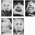
The serratus anterior muscle has been most useful in situations where a small, relatively thin, and potentially functional muscle is needed. As a free transfer, one or two slips are ideal for use in the hand or foot (4). The segmental organization allows careful axial division between slips for placement of portions of the muscle into various locations within a wound. Reanimation in facial paralysis requires three slips. In most cases, a cross-facial nerve graft is performed as a first stage. When axonal growth has advanced across the face, the lower three slips of the serratus can be inserted for functional replacement of the paralyzed muscle units (Fig. 155.1). With the muscle turned over, the neurovascular repairs are protected and only a short pedicle is required.
ANATOMY
The serratus anterior muscle is a broad, flat muscle that forms the medial wall of the axilla. Its dual blood supply and unique organization into separate slips that originate from each of the first nine ribs make it ideal for segmental transfer of only the lower three to four slips, thereby preserving muscle function (Fig. 155.2). The lateral thoracic artery vascularizes the upper four or five slips, whereas the lower portion is consistently supplied by a large branch of the thoracodorsal artery. This vessel enters the serratus in its posterior third and gives off segmental branches to each of the lower five slips (Fig. 155.3), each branch accompanied by a corresponding vein and a twig of the long thoracic nerve. The most inferior three slips are particularly suitable for use as a free muscle transfer and will have a long pedicle if the serratus branch is dissected back to include the subscapular-thoracodorsal axis (Fig. 155.4).
The serratus anterior stabilizes the scapula by its broad insertion along the medial and inferior border. It is innervated by the long thoracic nerve that arises from C5, C6, and C7 and travels inferiorly along the serratus fascia, joining with the serratus branch of the thoracodorsal artery at about the level of the sixth slip (Fig. 155.3). Loss of the long thoracic nerve will cause winging of the scapula, a complication that can be avoided by separating the discrete fascicles of the long thoracic nerve to only the lower three slips. The proximal serratus innervation must be preserved to maintain the muscle as a functional unit. With magnification, the long thoracic nerve can be split into fascicular groups to the level of the axillary vein, but this tedious exercise probably puts the proximal nerve at unnecessary risk of injury. Therefore, it is better to use a shorter nerve segment and to interpose a nerve graft at the recipient site if a long nerve pedicle is needed. Removal of the lowest three slips of the serratus will not result in winging of the scapula as long as the proximal muscle retains its innervation.
Related posts:


Stay updated, free articles. Join our Telegram channel

Full access? Get Clinical Tree


