Microneurovascular Free Gracilis Muscle Transfer for Facial Reanimation
K. HARII
H. ASATO
The free gracilis muscle transfer with microneurovascular anastomoses is currently a worthwhile and reliable procedure, especially in the successful reconstruction of a paralyzed face.
INDICATIONS
Since our clinical introduction of the free gracilis muscle transfer with microneurovascular anastomoses in 1976 (1), this procedure has provided many advantages for successful facial reanimation in long-standing or established facial paralysis. This procedure is indicated for a complete paralysis without regard to etiology. It can also be used for incomplete or partial paralysis.
The motor nerve of the gracilis muscle can be sutured to either the ipsilateral facial nerve or the stump of a previously performed cross-facial nerve graft (2). On some occasions, a branch of the hypoglossal nerve can be used.
ANATOMY
The gracilis muscle is the most superficially located muscle in the thigh adductor group. It arises from the medial margin of the lower half of the pubic arch and inserts into the medial surface of the upper end of the tibia. It forms a flat and narrow muscle belly.
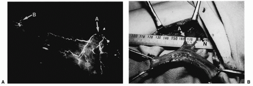 FIGURE 153.1 A: Vascular supply to the gracilis muscle. Arrow A, dominant nutrient vessels. Arrow B, small additional nutrient vessels. B: Elevated gracilis muscle. Arrow A, pedicle vessels. Arrow N, motor nerve. |
The dominant nutrient vessels of the gracilis muscle originate from either the profunda femoris or the medial circumflex femoral vessels and enter the muscle from its upper third. Although there are a few additional small nutrient vessels ramifying directly from the femoral vessels to the distal portion of the muscle, the dominant nutrient vessels alone can nourish the entire muscle belly (Fig. 153.1A).
The motor nerve of the gracilis muscle is derived from the anterior branch of the obturator nerve with the sensory nerve to the thigh, which separates from the motor nerve at the gracilis hilus and finally terminates at the skin of the inner medial thigh. The motor nerve of the gracilis muscle is accompanied by the dominant nutrient vessels and can easily be identified between the adductor longus and brevis muscles (Fig. 153.1B).
OPERATIVE TECHNIQUE
Isolation of the Muscle
An incision approximately 10 cm long is placed along the posterior border of the adductor muscle. After separation of the fascia over the adductor longus and gracilis muscles, the dominant nutrient pedicle of the gracilis is normally identified
between the adductor longus and brevis muscles. Blunt careful finger dissection around the muscles can easily expose the pedicle vessels and nerve without injury.
between the adductor longus and brevis muscles. Blunt careful finger dissection around the muscles can easily expose the pedicle vessels and nerve without injury.
Related posts:


Stay updated, free articles. Join our Telegram channel

Full access? Get Clinical Tree


