This article describes the indications for and methods of managing phalangeal fractures. The fractures around the finger joints are particularly difficult to treat. The technical details and pitfalls for these cases are discussed in detail. The problems associated with the phalangeal fractures such as articular destruction, nonunion, and postoperative care are also discussed. The authors present their preferred surgical treatment, with review of recent advancement regarding the treatment of phalangeal fractures.
Key points
- •
Stable reduction and joint alignment are the key tenants to fracture fixation.
- •
In the presence of severe soft-tissue (eg, skin, nerve, tendon, nail) injury, the outcome of phalangeal fractures can be difficult to optimize.
- •
Early joint motion needs to be initiated with a good balance with a stable construct; this is especially true of phalangeal fractures around the proximal interphalangeal (PIP) joint.
- •
The fractures involving the PIP joint are often associated with dislocation of the joint and are particularly difficult to treat.
- •
Early digital motion with severe soft-tissue damage, high velocity or blast, multidigit or severe fracture communition is often compromised.
- •
Postinjury rehabilitation plays a critical role and is an integral part of treatment.
Introduction
The phalanges of the hand are tubular structures. Proximal and distal interphalangeal (DIP) joints allow for predominantly flexion and extension. The collateral ligaments stabilize the joints and restrain their lateral motion. The volar plate and extensor apparatus further stabilize these joints.
During the last 20 years, although many conventional methods are still used for treatment of phalangeal fractures, one has seen the evolution of much lower-profile plate and screw implants, which improved outcomes. In addition, most surgeons favor less-invasive operative approaches. However, there are ongoing debates with regard to plate superiority over simpler, less-invasive methods of fixation, given the potential for adhesions. Exciting new fixation capabilities are emerging, such as percutaneous compression wire fixation and bioabsorbable implants for plate and screw fixation. Novel methods for managing common problems have been described, such as the bone peg for distal phalangeal nonunions and the potential use of bone graft substitutes. Aggressive early motion has been an important focal point for management of problematic injuries as well.
Evaluation and selection of treatment options
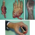
The history and physical examination are necessary for management decision making. Special attention should be paid to the mechanism, handedness, smoking history, comorbid disease precluding surgery, premorbid functionality of the patient, occupation, hobbies, and the ability to comply with the intended treatment program. A minor traumatic event resulting in a significant fracture should raise the suspicion of a potential pathologic condition such as a tumor or osteoporosis ( Fig. 1 ).
Patients with a significant smoking history are more likely to have delayed union or nonunion than nonsmokers. Patients with severe dementia and a displaced phalanx fracture may benefit from conservative intervention. Their functional demands and compliance with complex hand therapy often is suboptimal. Finally, the noncompliant patient does a disservice to both themselves and the surgery performed. Although it may be impossible to predict, a careful history or past experience may guide the surgeon in fixation method or choice in this difficult population.
Indications for conservative treatment include the following:
- 1.
Closed (or open) stable fracture patterns with adequate reduction
- 2.
Fractures with suboptimal reduction, good longitudinal alignment, with or without some shortening, but the fractures do not involve the joint and there is no rotational deformity.
- 3.
Patients unable to comply with surgical treatment because of their age or systemic or local wound conditions, or patients with much lower functional demands
- 4.
Noncompliant patients
Indications for surgical treatment include the following:
- 1.
Fractures that are persistently unstable after reduction. These fractures possess unacceptable shortening, rotation, angulation, or articular step-off
- 2.
Inability to obtain satisfactory fracture reduction with unacceptable shortening rotation, angulation, or articular step-off (>1 mm)
- 3.
Open fractures with severe soft-tissue injuries, and the fractures are unstable
- 4.
Fractures with displaced articular surface involvement, especially with multiple fragments, which cannot be reduced adequately
Treatment of phalangeal fractures: general guidelines
Fractures in Adults
Nonoperative treatment
Nonoperative treatment of phalangeal fractures is often managed with cast or splint immobilization for up to 4 weeks. Distal phalangeal fractures can be managed with Alumafoam (Hartmann, Rock Hill, SC) or custom Orthoplast (Patterson Medical, Warrenville, IL) splints. Elderly patients may need more prolonged immobilization, but stiffness easily develops. For nondisplaced fractures or stable fractures, buddy taping is an alternative method. Buddy taping can also be applied to those patients who have been treated with splint or cast fixation for the initial 2 weeks. Buddy taping allows for finger motion, which decreases the likelihood of joint stiffness.
Operative treatment
For displaced and unstable fractures, we prefer closed reduction and percutaneous fixation with 2 crossing Kirschner wires (K-wires). The closed K-wire placement does not involve periosteal stripping and decreases the chance of associated tendon adherence and damage to bone vascularity. If adequate reduction and stabilization is achieved, immediate gentle range of motion is commenced within a week of surgery. K-wire removal and progression of therapy is determined by clinical examination, usually within 4 weeks.
Open reduction is warranted in fractures that are not reducible by closed means, open fractures with concomitant nerve or tendon injury, and fractures with significant bone loss. Some intra-articular fracture may require open reduction to reduce the fracture or to restore smooth articular surface. Open reduction and fixation with K-wires, low-profile titanium plates, lag screws, cerclage wires, or dynamic external fixators allow at least some extent of early motion, which prevents finger joint stiffness and tendon adherence to the fracture site or hardware. It is important to inform the patients about risks of potential complications such as infection, malunion, nonunion, arthritis, hardware complications, stiffness, and tendon adhesions. Specific fracture patterns, with focus on problematic injuries, are discussed in the subsequent sections.
Pediatric Fractures
Pediatric fractures can heal quickly and are often healed in a short period (about or less than 3 weeks). This quick healing is advantageous. However, pediatric patients must be protected with a cast or a splint for more than 4 weeks because they are more active.
In addition, the remodeling potential of pediatric fractures is great; these fractures tolerate a greater degree of angular deformation, as the deformation tends to disappear during growth. Overall, these fractures demonstrate a greater degree of remodeling capacity, allowing for more conservative management in many instances. We believe that absolute surgical indications for a pediatric fracture include open fractures with significant comminution or displacement. Fractures that involve joint surfaces with considerable malalignment also require operative intervention. Closed fractures that cannot be reduced in a closed manner, with significant displacement, concern for soft-tissue entrapment, or the presence of scissoring, are indications for surgical management.
Also, unique to skeletally immature fractures are injuries that involve physis. The most commonly used classification system for these types of fractures is that of Salter-Harris. This classification ranges from I to IX, with the most common injuries in the range from I to V ( Table 1 ).
| Type | Salter-Harris Classification |
|---|---|
| I | A fracture that involves the physis |
| II | A fracture that involves the physis and metaphysis; this is the most common fracture pattern |
| III | A fracture that involves the physis and epiphysis |
| IV | A fracture that involves the metaphysis, physis, and epiphysis |
| V | A compressive or crush-type injury to the epiphyseal plate |
| VI | An injury to the perichondrial structures |
| VII | An isolated injury to the epiphyseal plate |
| VIII | An injury that is isolated to the metaphysis, there may be a potential for enchondral ossification |
| IX | An injury to the periosteum that may interfere with normal membranous growth |
There is a subset of pediatric injuries that may be particularly problematic if not managed appropriately. This subset includes phalangeal neck fractures, intercondylar fractures, Seymour fractures (an extra-articular transverse fracture of the base of the distal phalanx with flexion deformity at the fracture site), and Salter-Harris III physeal fractures. These fractures necessitate appropriate reduction and close monitoring. Malunion occurs more often with all these fractures. However, nonunion and avascular necrosis, although extremely rare, may occur in the pediatric hand population. In one series reported by Al-Qattan, 4 (2%) digits of 215 phalangeal neck fractures in children developed nonunion, predominantly in the proximal phalanx of the thumb. Eight (4%) patients had nonunion with avascular necrosis in the proximal phalanx of the small finger, all in older children.
Distal phalanx fractures
Common fracture patterns of the distal phalanx include tuft fractures, shaft fractures, and articular fractures ( Table 2 ). Most commonly, these injuries are a result of a crushing force to the fingertip in both adults and children.
| Tuft Fractures | Shaft Fractures | Articular Fractures |
|---|---|---|
| Simple | Transverse | Dorsal (mallet) fractures |
| Comminuted | Oblique | Volar (jersey) fracture |
| Longitudinal | Epiphyseal/Salter-Harris | |
| Subtypes: stable/unstable/comminuted |
Tuft Fractures
A short period of immobilization, 2 to 4 weeks, is often adequate for tuft fractures and about 4 weeks for minimally displaced shaft fractures. Tuft fractures may not seem to have full osseous union on follow-up radiographs but often have an adequate stable fibrous union. Minimally displaced or stable-after-reduction shaft fractures of the distal phalanx are immobilized with Alumafoam (Hartmann) splint or custom Orthoplast (Patterson Medical) splint. The DIP joint is included in the splint. Patients are instructed in the maintenance of suppleness of noninvolved joints to avoid stiffness.
Displaced, unstable shaft fractures or painful tuft fractures with delayed union ( Fig. 2 ) may necessitate reduction and internal splinting with K-wires along with nail matrix repair (if indicated). Using fluoroscopy, K-wires are driven in a retrograde fashion from the distal hyponychium just under the nail plate to the subchondral distal phalanx not crossing the DIP joint. One or two K-wires may be indicated to control rotation. Our preferred method is to use one 0.045-in K-wire or a 0.045-in wire combined with a 0.035-in wire. These wires are cut just under the skin, and a single suture is placed after irrigation of the wound. Alternatively, the wire can be left out of the skin and bent; a wire protector is placed to prevent inadvertent injury. The advantage of burying the wire is hygiene, decreased infection rate, and ability to get the finger wet; the disadvantage is the effort of removal.
Other methods for intraosseous fixation are cortical screws or variable pitch compression screws. These screws can be used in the management of symptomatic nonunions as well. Bone grafting may be necessary for nonunion with dorsal or palmar approach. If stable fixation is achieved with 2 wires, cortical screws, or variable pitch screws not crossing the DIP joint, then the patient wears a removable splint, which can be removed intermittently during the day to perform active DIP joint motion exercises. Removal of K-wires ensues once clinical evidence of bony healing has been achieved usually at 4 weeks. Loading of the joint and pinching exercises can then progress.
Intraarticular Fractures of the DIP Joint
Periarticular injuries of the DIP joint can present with varying complexity. Bony mallet injuries result from forced hyperflexion of the DIP joint, with disruption of the terminal extensor tendon and attached bony articular fragment. These injuries can often be managed conservatively with extension splinting of the DIP joint. However, significant joint involvement, greater than a third articular surface, with subluxation of the remainder of the phalanx in a volar direction warrants surgical intervention. Achievement of joint congruity is obligatory to avoid stiffness and arthritis from malunion of the DIP joint with closed or open treatment. Closed options include reduction with extension block pinning and/or transarticular pinning. Open reduction and pin fixation, mini screws, tension band, hook plates, pullout wires/button, or external fixation are also surgical options.
Our preference in these unstable mallet fracture dislocations is to perform extension block and transarticular K-wire fixation.
- 1.
The DIP joint is maximally flexed, and a 0.045-in K-wire is driven at a 45° angle into the dorsal head of the middle phalanx to buttress against the dorsal articular fragment.
- 2.
The fracture is reduced, the finger extended, and another 0.045-in K-wire is placed percutaneously in a retrograde fashion from the fingertip pinning the DIP joint.
- 3.
If the dorsal articular fragment was large enough, then interfragmentary fixation can be performed with another 0.045 or 0.035-in K-wire or opened as described previously.
The ideal method has not been elucidated; multiple investigators recommend open reduction internal fixation (ORIF), whereas others report higher complication rate and stiffness with open technique and thus espouse closed management. Hofmeister and colleagues retrospectively reviewed 24 acute and chronic bony mallet injuries treated with extension block pinning and transarticular fixation. Their indications for surgical management included closed displaced mallet fractures with greater than 25% involvement of the joint surface; the average fracture size of their cases was 40% of the articular surface. They removed the pin at 4 weeks, followed by volar splinting for 2 weeks while allowing extension exercises during this period. The time to union was on average 35 days. The fractures had no major complications, and their average extension loss was 4° with flexion 77°. Lucchina and colleagues reviewed 58 acute mallet fractures with greater than a third joint involvement with volar subluxation of the distal fragment, using extension block pinning, K-wires used as joysticks, and ORIF with interfragmentary mini screws. They found that K-wires were more technically facile compared with the technically demanding ORIF. However, internal fixation allowed earlier mobilization and return to work.
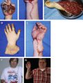
The bony jersey finger is another type of injury. It is a volar articular fracture secondary to avulsion of the flexor digitorum profundus, with an associated bony fragment. These injuries are uncommon and usually require surgical management with ORIF of larger fragments with mini screws. Smaller fragments require tendon fixation to bone with suture anchor or pullout wire/button, usually within 10 days of injury as the flexor retracts ( Fig. 3 ). In our experience with small fragments, the flexor digitorum profundus needs to be retrieved in the palm and fixated to bone with mini anchors or pullout suture with button. For larger fragments with attached tendon, articular congruity is reestablished with dorsal block splinting if stable or with ORIF of the fragment with screws, K-wires, or pullout wire.
Malreduction of pediatric extra-articular transverse physeal or juxtaepiphyseal fractures of the distal phalanx (ie, Seymour fractures) often go unrecognized. This fracture is a result of a hyperflexion injury. The extensor maintains insertion on the proximal epiphysis and the flexor digitorum profundus on the distal fragment, resulting in dorsal apex configuration and fracture through the nail bed. Simple reduction maneuvers can result in inadequate reduction and nail bed entrapment. Infection, malreduction, permanent bump, poor motion, and distal phalangeal shortening are concerns with inadequately treated injuries. Treatment should consist of irrigation, reduction of the nail bed and fracture, repair of the nail bed, nail plate stenting, and extension splinting. These injuries are traditionally stable after nail bed repair and stenting, but pin fixation may be considered for stabilization if warranted ( Fig. 4 ).
On review of our patients during a 5-year period, most nonoperative tuft fractures went on to heal uneventfully. The preponderance of patients, adult and children, sustained crush injuries from doors or other mechanisms. The remainder occurred in adults predominantly from machines such as table saws, with a mixture of open tuft, shaft, and periarticular injuries.
In our series of 43 operative distal phalangeal fractures, only 2 were tuft fractures. One patient required excision of a painful distal tuft remnant, and one required K-wire fixation of a distal tuft nonunion. All patients requiring fixation were treated with K-wires. Distal phalangeal shaft fractures, albeit a small percentage of all distal phalanx fractures, were more commonly unstable, warranting percutaneous K-wire fixation for stabilization. Articular fractures were more likely to necessitate open reduction than shaft fractures, but the majority were treated percutaneously. All patients were pain-free by 6 months, with the majority back to work within 6 weeks for nonarticular fractures. Range of active motion approximated 60° for nonarticular injuries. Patients with articular fractures suffered from the least regain of motion. The common complication outside of loss of range of active motion was nail deformity.
Complications
All phalangeal fractures have the potential to develop nonunion, malunion, infection, and poor DIP joint motion. Specific concerns for the distal phalanx include infection, nail bed deformity, growth delay for pediatric patients, and painful nonunions. If indicated, most painful delayed nonunions are treated with open debridement, bone grafting, and stabilization with K-wires, cortical screws, or cannulated variable pitch compression screws. Articular fractures with poor motion and pain may require later arthrodesis. One point that should not be minimized is the degree of soft-tissue injury associated with these fractures. With severe soft-tissue injury, the complication rate should be expected to be higher. Management of patient expectations along with the associated complication is necessary.
Proximal and middle phalanx fractures
Fractures of the middle and proximal phalanges are initially divided into articular and nonarticular. Fractures involving the joint are then further broken down into anatomic location, such as condylar (uni or bicondylar), shape (oblique, volar, dorsal, sagittal, or coronal), and whether there is comminution present. Similarly, fractures that do not involve the joint can be further classified based on location (neck, shaft, or metaphyseal), shape (oblique, spiral, or transverse), and finally whether comminution is present.
Volar, dorsal, and pilon fracture dislocations of the base of the middle phalanx deserve special mention, as these are unstable with subluxation of articular fragments. Significant disability with regard to stiffness and pain can occur. Frequently, early operative intervention is indicated.
Fractures That Involve the Joint
It is expected that fractures that involve the PIP and DIP joints will have some degree of postinjury stiffness and possible motion abnormality. Ideally, maintaining motion while using a stable method of fixation during rehabilitation would provide the best outcome. This method is not always possible. Displaced and unstable intraarticular fractures warrant surgical treatment to realign the osteochondral fragments. This treatment is quite challenging with regard to fixation given the small fragments often involved. Most investigators argue that surgical management is necessary in these displaced fractures given the poor outcomes of nonoperative treatment with associated pain, stiffness, and late secondary osteoarthritis. Surgical management and the complications associated with these troublesome injuries are discussed.
Phalangeal head fractures may be unicondylar or bicondylar, displaced or nondisplaced. The collateral ligaments are traditionally associated with the fracture fragments. If nondisplaced, buddy taping and a therapy program may suffice. As these fractures are inherently unstable, close follow-up with weekly radiologic examination is necessary. Displacement can occur in a delayed fashion. If displaced, reduction is required for these injuries. Fixation techniques that may be used are K-wires, lag screws, cannulated mini screws, interosseous wiring/tension bands, and unicondylar plating. If screws are used, the smaller 1.0- to 1.5-mm screws are appropriate. It is important to remember that single-point fracture fixation with K-wires is not a stable technique, and the fracture may displace with time. If there is insufficient bone fragment remnant for extra-articular screw placement, intraarticular fixation with countersinking may be required. Surgical approach can be midaxial, bilateral midaxial, or dorsal curvilinear depending on the fracture type.
Bicondylar fractures
Bicondylar fractures are extremely challenging. The amount of bone stock present and the size of the fragment often dictate the fixation method. Standard fixation techniques noted in the unicondylar fracture pattern may be used to stabilize a bicondylar fracture. Anatomic reduction of bicondylar fractures is even more difficult in closed fashion. The achievement of articular congruity likely requires open methods. Our method is to first rebuild the head of the phalanx by fixating the condylar segments together either with mini screws or K-wires, followed by fixating the construct to the remainder of the phalanx with K-wires. This procedure can be performed with a variety of fixation strategies as described earlier ( Fig. 5 ).