44
Metacarpal Head Fractures
Paul R. Greenlaw and Mark R. Belsky
History and Clinical Presentation
A 58-year-old right hand dominant woman presented for evaluation of an injury to the left hand. The patient had fallen on her left hand the previous day. Overnight she noted severe swelling in the left hand, which was treated with ice and ibuprofen. She complained of only left hand pain and denied paresthesias or paralysis in the left hand at presentation.
Physical Examination
The patient’s pertinent findings were confined to the left hand. Swelling and tenderness with intact skin were noted around the heads of the fourth and fifth metacarpals of the left hand. Range of motion of the fourth and fifth metacarpophalangeal (MP) joints was limited secondary to pain. Sensation and circulation were intact in the left ring and small finger with adequate alignment and rotation of the digits. No collateral ligament instability was appreciated.
Diagnostic Studies
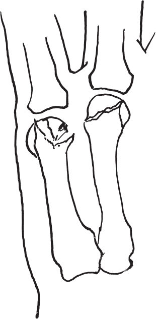
Anteroposterior, lateral, and oblique radiographs of the left hand (Fig. 44–1) revealed a nondisplaced shear fracture of the fourth metacarpal head and a displaced sagittal fracture of the head of the fifth metacarpal extending into the metacarpal neck.
Differential Diagnosis
Fracture
Intraarticular
Extraarticular
Sprain
PEARLS
- The goal of ORIF is to obtain anatomic reduction of the joint surface with fixation that will allow early motion.
PITFALLS
- Small fragments require meticulous handling to avoid devascularization and resultant avascular necrosis.
Diagnosis
Sagittally Oriented Fifth Metacarpal Head Fracture with Extension into the Fifth Metacarpal Neck and Nondisplaced Shear Fracture of the Fourth Metacarpal Head
Metacarpal head fractures are uncommon fractures. A comprehensive review by McEl-fresh and Dobyns revealed an array of metacarpal head fracture patterns they classified into 10 types based on anatomic involvement as seen on roentgenographic examination: (1) epiphyseal, (2) collateral ligament avulsion injuries, (3) osteochondral, (4) oblique (sagittal), (5) vertical (coronal), (6) horizontal (transverse), (7) comminuted, (8) intraarticular extension (in boxers), (9) loss of substance, and (10) avascular necrosis. Of these types, comminuted fractures were the most common (31%).


Figure 44–2. A stable fourth metacarpal head fracture and displaced fracture of the head of the fifth metacarpal.
Surgical Management
Stay updated, free articles. Join our Telegram channel

Full access? Get Clinical Tree


