11
Mandibular Fractures
 Anatomy
• A “U-” shaped bone that contains two hemimandibles
• Structures unite at midline called symphysis
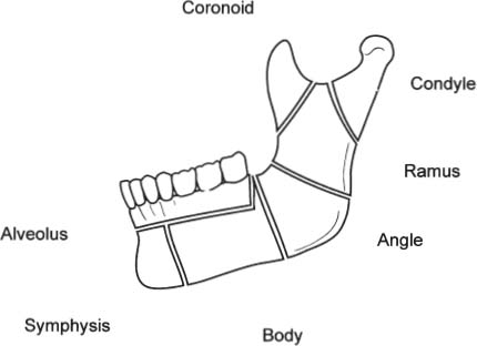
• Each hemimandible consists of a (Fig. 11–1)
 Body
Body
 Angle
Angle
 Ramus
Ramus
 Coronoid process
 Condyle
• Muscles of mastication
 Jaw protrusion
 Lateral pterygoid (lateral pterygoid plate to condylar neck)
 Jaw elevators
 Temporalis (temporal fossa to coronoid)
 Masseter (zygomatic arch to the body)
 Medial pterygoid (medial pterygoid plate to angle)
 Jaw depressor-retractors
 Lateral pterygoid
 Digastric
 Geniohyoid
 Mylohyoid
 Genioglossus

Figure 11–1 Anatomy of the mandible.
• Condyle articulates with cranium at the glenoid fossa of the temporomandibular joint (TMJ)
• Blood supply of mandible
 Inferior alveolar artery from the internal maxillary artery enters at mandibular foramen and exits at mental foramen
 Branches from the muscles of mastication
• Nerve supply
 Inferior alveolar nerve from V3 enters at mandibular foramen and exits at mental foramen
• Mental foramen
 Located between first and second premolar
 Dental Relationships
Child:
• 20 deciduous or primary teeth labeled A – T
 Right A B C D E F G H I J
 Left T S R Q P O N M L K
Adult:
• 32 permanent teeth labeled 1 through 32
 Numbering begins with the third right maxillary molar as tooth #1 and the last maxillary molar as #16
 Numbering continues onto the mandibular left third molar as #17 and ends with the mandibular right third molar as #32
Each hemimandible or hemimaxilla consists of
• One central and one lateral incisor
• One canine (cuspid)
• First and second premolar (bicuspid)
• First, second, and third molar
Angle Classification of Occlusion
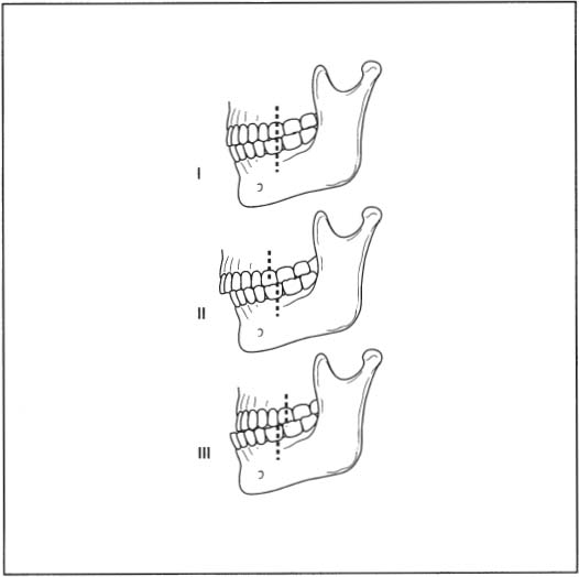
Based on the first maxillary molar and its position to the first mandibular molar (Fig. 11–2):

Figure 11–2 Angle classification of occlusion.
• Class I – normal occlusion
 Mesiobuccal cusp of the maxillary first molar occludes with buccal groove of the mandibular first molar
• Class II – overbite
 Lower first molar is distal (posterior) to the upper first molar
• Class III – underbite
 Lower first molar is mesial (anterior) to the upper first molar
 Mandibular Fractures
Stay updated, free articles. Join our Telegram channel

Full access? Get Clinical Tree


