Capillary malformations (CMs) are the most common vascular malformations. They are comprised of the small vessels of the capillary network in skin and mucous membranes. In the vast majority of affected individuals, CMs are isolated and not associated with any underlying abnormalities. Depending on size and location, however, they may cause significant morbidity due to disfigurement or stigmatization and, rarely, herald the presence of an underlying syndrome.
Capillary malformations (CMs) are the most common vascular malformations. They are comprised of the small vessels of the capillary network in skin and mucous membranes. In the vast majority of affected individuals, CMs are isolated and not associated with any underlying abnormalities. Depending on size and location, however, they may cause significant morbidity due to stigmatization or disfigurement and, rarely, herald the presence of an underlying syndrome.
Fading capillary stain (nevus simplex)
The fading capillary stain, also known as salmon patch or stork bite, is the most common vascular stain, present in up to 40% of infants. It consists of dilated capillaries within the papillary dermis. Some postulate that they are due to lack of autonomic regulation of local vessels in the affected skin. In general, fading capillary stains on the face diminish over time and those on the nape of neck tend to persist. Fading capillary stains are distinct from CMs and other vascular malformations. They are often observed on the glabella, eyelids, nose, and philtrum and even more frequently seen on the posterior neck. The lumbosacral spine can also be affected and some clinicians think that in this location it can rarely be associated with occult spinal dysraphism. Many infants have stains on multiple sites. Unlike true CMs, fading capillary stains on the face do not occur in a dermatomal distribution and are not associated with the Sturge-Weber syndrome (SWS). Large and persistent stains on the face, however, are sometimes seen in association with other underlying diseases or syndromes, such as the Beckwith-Wiedemann syndrome and macrocephaly-CM syndrome (M-CM).
Usually, no treatment is required. In persistent stains on cosmetically sensitive locations (ie, facial), however, treatment with the pulsed dye laser (PDL) is effective and can be considered. In infants and children with persistent stains, a detailed history and physical examination to rule out associated abnormalities, such as macrocephaly, hypotonia, or other clinical features seen in M-CM and Beckwith-Wiedemann syndrome, could help facilitate the diagnosis of one of these conditions. In patients with lumbosacral stains, particularly those with the reportedly characteristic butterfly shape, it may be appropriate to consider imaging studies, such as duplex ultrasound or MRI, to rule out underlying occult spinal dysraphism. This is controversial, however, and prospective studies are needed to determine the true association, which is probably low.
Capillary malformations
CM is equated with port wine stain, which is what it is still most commonly referred to in the literature. CM is also known as nevus flammeus. CM is common and occurs in approximately 0.3% of all newborns. Histolopathologically, initially CM is composed of normal capillaries in the superficial dermis with no evidence of cellular proliferation. Telangiectasias and vascular ectasias become more evident over time. CMs are true vascular malformations and as such are present at birth and persist throughout life. They can occur anywhere on the body. Growth is proportionate with that of the affected child. On the face, they usually present in a dermatomal distribution and respect the midline. In some cases, however, there is involvement of neighboring dermatomes. CMs are initially bright pink, red, or violaceous in color. They often seem to lighten significantly over the first few months of life, probably due to a drop in circulating blood hemoglobin concentration. This physiologic lightening is not indicative of spontaneous resolution. Most CMs become darker, thicker, and more nodular over time, particularly within facial lesions. Local complications of CM include the development of pyogenic granulomas and eczematous dermatitis occurring within the stain. The differential diagnosis of CM includes a host of other vascular lesions, both tumors and malformations, including fading capillary stain, early infantile hemangioma, telangiectatic or nonproliferative infantile hemangioma, vascular stain associated with arteriovenous malformation (AVM), and other vascular malformations, including lymphatic and/or combined malformations (ie, capillary-lymphatic-venous malformation [CLVM]). Early in infancy it can sometimes be difficult to distinguish between CM and infantile hemangiomas located on the face. Clues to the diagnosis include the distribution (dermatomal in CM versus regional or segmental in infantile hemangiomas) or the presence of small, brightly erythematous islands of proliferation characteristic of infantile hemangioma ( Fig. 1 ). To differentiate between CM and underlying AVM, Doppler assessment of flow can be helpful along with auscultation for a bruit and palpation for the thrill of an AVM.
Associated Syndromes
Inherent in the management of infants and children with CM is the initial evaluation and continued monitoring for associated syndromes. Although rare, CM can present as manifestations of several distinct syndromes. In the past, many different vascular malformations were confused with CM; however, since the advent of the International Society for the Study of Vascular Anomalies (ISSVA) and the development of a specific and updated classification framework for vascular lesions, vascular malformations can now be more precisely identified and separated into their respective vessels of origin. Syndromes that exhibit true CM include SWS (which is the most common), M-CM, capillary malformation-arteriovenous malformation syndrome (CM-AVM), cutis marmorata telangiectatica congenita (CMTC), and overgrowth syndromes, such as Klippel-Trénaunay syndrome (KT). Several of these entities, including KT and CM-AVM, are described in subsequent articles.
Sturge-Weber syndrome, OMIM 185300
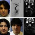
SWS refers to the triad of a facial CM in the V1 distribution, ipsilateral leptomeningeal vascular malformation, and choroidal vascular malformation of the eye, which can lead to ipsilateral glaucoma and buphthalmos. The cause is unknown; cases are sporadic with no clear evidence of genetic predisposition.
Approximately 6% to 10% of patients with a CM in the V1 distribution have SWS. Bilateral involvement and involvement of the other trigeminal dermatomes may also occur and increase the risk of central nervous system (CNS) involvement. There does not seem to be any direct relationship between the size of the CM and the severity of the brain involvement in affected patients. Common neurologic manifestations include contralateral seizures, hemiparesis or hemiplegia, migraine headaches, and intellectual impairment. Seizures occur in 75% of patients; these are usually generalized tonic-clonic type and onset is usually before age 1. Developmental delay occurs in approximately half of affected children and prolonged or uncontrolled seizures can worsen CNS outcomes (ie, poor intellectual development, behavioral abnormalities, and learning disabilities). Aggressive seizure management is often warranted.
The incidence of glaucoma in association with V1 CM has been reported as between 7% and 52%. Like CNS involvement, the incidence of glaucoma is much higher when V1 and V2 dermatomes are involved. The presence of a CM in the V2 distribution has not been associated with the development of glaucoma. Regular ophthalmologic examinations (every 6 to 12 months) should be performed in all affected patients, because glaucoma can develop early. Early detection of raised intraocular pressure is important in preventing progressive disease. Long-term follow-up is also necessary because glaucoma can arise as a late complication. When glaucoma is present early on, associated bupthalmos (bull’s eye/large eye secondary to enlarged cornea) can also occur. Diffuse vascular malformation of the choroid ipsilateral to the CM is another characteristic feature of SWS that may be found in up to 71% of SWS cases. Myopia, coloboma, cataract, and iris heterochromia have also been seen in association with SWS.
Children with facial CM in the V1 or multiple dermatomal distributions should be evaluated for possible SWS. A complete fundoscopic eye examination and evaluation of intraocular pressure should be performed at regular intervals. In those patients with known eye complications, early-onset seizures, or other symptoms, neuroimaging to identify leptomeningeal (pial) vascular malformations should be performed. MRI with gadolinium contrast is the most sensitive study for diagnosis and evaluation of SWS. In addition to the ipsalateral vascular malformations, cerebral cortex atrophy or characteristic calcifications can also often observed in affected patients.
The management of SWS depends on the clinical manifestations. Seizures require aggressive anticonvulsant therapy and regular follow-up with neurology. In very severe, refractory cases, surgical intervention, such as localized resection of the involved brain tissue or hemispherectomy, may be warranted. In some cases, early neurosurgical intervention may improve outcomes.
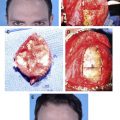
Laser treatment of the CM can begin early (ie, when seizures are controlled by anticonvulsants). Patients often require many treatments and even repeated laser ablation (discussed later) years later because CMs are known to undergo redarkening. Many patients with SWS have associated soft tissue and bony overgrowth of the affected areas. Gum hypertrophy, a common finding in patients with V2 involvement and overgrowth of the maxilla, is exacerbated by antiepileptic medications but can be amenable to surgical resection. The lip and surrounding soft tissue hypertrophy are the most common sites requiring surgical intervention. Overgrowth of the maxilla creates an occlusion deformity and crossbite requiring regular orthodontic evaluation and follow-up. Patients with SWS benefit from a multidisciplinary approach with the expertise of neurologists, ophthalmologists, pediatricians, neurosurgeons, dermatologists, plastic surgeons, maxillofacial surgeons, otolaryngologists, and neuroradiologists. The Sturge-Weber Foundation is an active support group for patients and families ( http://www.sturge-weber.org/ ).
Macrocephaly–capillary malformation, OMIM 602501
M-CM was previously known as macrocephaly-CMTC (M-CMTC). Recently more appropriately named, M-CM is a rare, sporadic syndrome characterized by either CM or persistent nevus simplex with macrocephaly, hypotonia, and developmental delay. The vascular stain of M-CM is characteristically ill defined or blotchy and is not be confused with true CMTC because it lacks the fixed livedoid pattern, atrophy, or ulceration, which can occur with CMTC. In M-CM, the vascular stain is usually located on the face; the philtrum and glabella are often involved. Other associated features include hydrocephalus, seizures, developmental delay, connective tissue defects (soft skin and joint hypermobility), toe syndactyly, frontal bossing, and, rarely, hemihypertrophy.
Capillary malformation–arteriovenous malformation, OMIM 608354
CM-AVM is an autosomal dominant condition recently found to be due to underlying mutations in the RASA1 gene (RASA1 is an inhibitor of the Ras-Map kinase pathway). The disorder is characterized by small, multifocal CMs in association with underlying AVMs/arteriovenous fistulas and occasionally Parkes Weber syndome. Affected patients demonstrate atypical-appearing CM, manifesting as randomly distributed, multifocal, pink to dull red annular papules/plaques with a surrounding halo of vasoconstriction. The lesions often demonstrate high flow or a bruit on doppler examination. In patients with multifocal CM, a Doppler examination is helpful to determine if there is an underlying AVM. Genetic counseling may be indicated.
Cutis marmorata telangiectatica congenita
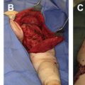
CMTC is an unusual, distinctive vascular malformation usually noted at birth. The cause is unknown and there is no known genetic basis. Infants present with a fixed coarse, violaceous, livedoid (or net-like) pattern of vasculature, which is unresponsive to local warming. Most commonly, these malformations occur in a regional distribution and respect the midline. Usually, the extremitites are involved ( Fig. 2 ). The affected limb may demonstrate hyper- or hypoplasia. Limb asymmetry is the most common associated finding (33%–68%), but lesions can also present with cutaneous atrophy or ulceration.
Neurologic complications, syndactyly, arterial stenosis, and ophthalmologic anomalies have also been reported, although less frequently. Histopathologically, CMTC consists of dilated capillaries and venules along with occasional atrophy and ulceration. The livedoid stains tend to improve in appearance over time. Follow-up with orthopedics is important when limb length discrepency is a concern.
Klippel Trenaunay, OMIM 149000
Traditionally, KT has been characterized as the triad of CM, visible venous varicosities, and bony and/or soft tissue overgrowth. It has become accepted, however, that the vascular malformation associated with true KT is actually a complex, combined vascular malformation (CLVM) associated with bony and soft tissue overgrowth of the affected limb. In most patients with KT, the CLVM presents as a well-demarcated, geographic violaceous plaque, often with visible nodules (lymphatic blebs), which become thicker over time.
Reticulate or blotchy CM on the extremities can also be associated with overgrowth and venous varicosities but are less likely to demonstrate extensive underlying lymphatic malformation. These patients with diffuse or blotchy CM of an extremity and stable overgrowth have a significantly better overall prognosis than those with true KT ( Fig. 3 ). In KT, progressive worsening of the venous stasis and/or lymphedema is inevitable, and ulceration, coagulopathy, thrombosis, pulmonary emboli, and pulmonary artery hypertension have all been associated.
Capillary malformations
CM is equated with port wine stain, which is what it is still most commonly referred to in the literature. CM is also known as nevus flammeus. CM is common and occurs in approximately 0.3% of all newborns. Histolopathologically, initially CM is composed of normal capillaries in the superficial dermis with no evidence of cellular proliferation. Telangiectasias and vascular ectasias become more evident over time. CMs are true vascular malformations and as such are present at birth and persist throughout life. They can occur anywhere on the body. Growth is proportionate with that of the affected child. On the face, they usually present in a dermatomal distribution and respect the midline. In some cases, however, there is involvement of neighboring dermatomes. CMs are initially bright pink, red, or violaceous in color. They often seem to lighten significantly over the first few months of life, probably due to a drop in circulating blood hemoglobin concentration. This physiologic lightening is not indicative of spontaneous resolution. Most CMs become darker, thicker, and more nodular over time, particularly within facial lesions. Local complications of CM include the development of pyogenic granulomas and eczematous dermatitis occurring within the stain. The differential diagnosis of CM includes a host of other vascular lesions, both tumors and malformations, including fading capillary stain, early infantile hemangioma, telangiectatic or nonproliferative infantile hemangioma, vascular stain associated with arteriovenous malformation (AVM), and other vascular malformations, including lymphatic and/or combined malformations (ie, capillary-lymphatic-venous malformation [CLVM]). Early in infancy it can sometimes be difficult to distinguish between CM and infantile hemangiomas located on the face. Clues to the diagnosis include the distribution (dermatomal in CM versus regional or segmental in infantile hemangiomas) or the presence of small, brightly erythematous islands of proliferation characteristic of infantile hemangioma ( Fig. 1 ). To differentiate between CM and underlying AVM, Doppler assessment of flow can be helpful along with auscultation for a bruit and palpation for the thrill of an AVM.
Associated Syndromes
Inherent in the management of infants and children with CM is the initial evaluation and continued monitoring for associated syndromes. Although rare, CM can present as manifestations of several distinct syndromes. In the past, many different vascular malformations were confused with CM; however, since the advent of the International Society for the Study of Vascular Anomalies (ISSVA) and the development of a specific and updated classification framework for vascular lesions, vascular malformations can now be more precisely identified and separated into their respective vessels of origin. Syndromes that exhibit true CM include SWS (which is the most common), M-CM, capillary malformation-arteriovenous malformation syndrome (CM-AVM), cutis marmorata telangiectatica congenita (CMTC), and overgrowth syndromes, such as Klippel-Trénaunay syndrome (KT). Several of these entities, including KT and CM-AVM, are described in subsequent articles.
Sturge-Weber syndrome, OMIM 185300
SWS refers to the triad of a facial CM in the V1 distribution, ipsilateral leptomeningeal vascular malformation, and choroidal vascular malformation of the eye, which can lead to ipsilateral glaucoma and buphthalmos. The cause is unknown; cases are sporadic with no clear evidence of genetic predisposition.
Approximately 6% to 10% of patients with a CM in the V1 distribution have SWS. Bilateral involvement and involvement of the other trigeminal dermatomes may also occur and increase the risk of central nervous system (CNS) involvement. There does not seem to be any direct relationship between the size of the CM and the severity of the brain involvement in affected patients. Common neurologic manifestations include contralateral seizures, hemiparesis or hemiplegia, migraine headaches, and intellectual impairment. Seizures occur in 75% of patients; these are usually generalized tonic-clonic type and onset is usually before age 1. Developmental delay occurs in approximately half of affected children and prolonged or uncontrolled seizures can worsen CNS outcomes (ie, poor intellectual development, behavioral abnormalities, and learning disabilities). Aggressive seizure management is often warranted.
The incidence of glaucoma in association with V1 CM has been reported as between 7% and 52%. Like CNS involvement, the incidence of glaucoma is much higher when V1 and V2 dermatomes are involved. The presence of a CM in the V2 distribution has not been associated with the development of glaucoma. Regular ophthalmologic examinations (every 6 to 12 months) should be performed in all affected patients, because glaucoma can develop early. Early detection of raised intraocular pressure is important in preventing progressive disease. Long-term follow-up is also necessary because glaucoma can arise as a late complication. When glaucoma is present early on, associated bupthalmos (bull’s eye/large eye secondary to enlarged cornea) can also occur. Diffuse vascular malformation of the choroid ipsilateral to the CM is another characteristic feature of SWS that may be found in up to 71% of SWS cases. Myopia, coloboma, cataract, and iris heterochromia have also been seen in association with SWS.
Children with facial CM in the V1 or multiple dermatomal distributions should be evaluated for possible SWS. A complete fundoscopic eye examination and evaluation of intraocular pressure should be performed at regular intervals. In those patients with known eye complications, early-onset seizures, or other symptoms, neuroimaging to identify leptomeningeal (pial) vascular malformations should be performed. MRI with gadolinium contrast is the most sensitive study for diagnosis and evaluation of SWS. In addition to the ipsalateral vascular malformations, cerebral cortex atrophy or characteristic calcifications can also often observed in affected patients.
The management of SWS depends on the clinical manifestations. Seizures require aggressive anticonvulsant therapy and regular follow-up with neurology. In very severe, refractory cases, surgical intervention, such as localized resection of the involved brain tissue or hemispherectomy, may be warranted. In some cases, early neurosurgical intervention may improve outcomes.
Laser treatment of the CM can begin early (ie, when seizures are controlled by anticonvulsants). Patients often require many treatments and even repeated laser ablation (discussed later) years later because CMs are known to undergo redarkening. Many patients with SWS have associated soft tissue and bony overgrowth of the affected areas. Gum hypertrophy, a common finding in patients with V2 involvement and overgrowth of the maxilla, is exacerbated by antiepileptic medications but can be amenable to surgical resection. The lip and surrounding soft tissue hypertrophy are the most common sites requiring surgical intervention. Overgrowth of the maxilla creates an occlusion deformity and crossbite requiring regular orthodontic evaluation and follow-up. Patients with SWS benefit from a multidisciplinary approach with the expertise of neurologists, ophthalmologists, pediatricians, neurosurgeons, dermatologists, plastic surgeons, maxillofacial surgeons, otolaryngologists, and neuroradiologists. The Sturge-Weber Foundation is an active support group for patients and families ( http://www.sturge-weber.org/ ).
Macrocephaly–capillary malformation, OMIM 602501
M-CM was previously known as macrocephaly-CMTC (M-CMTC). Recently more appropriately named, M-CM is a rare, sporadic syndrome characterized by either CM or persistent nevus simplex with macrocephaly, hypotonia, and developmental delay. The vascular stain of M-CM is characteristically ill defined or blotchy and is not be confused with true CMTC because it lacks the fixed livedoid pattern, atrophy, or ulceration, which can occur with CMTC. In M-CM, the vascular stain is usually located on the face; the philtrum and glabella are often involved. Other associated features include hydrocephalus, seizures, developmental delay, connective tissue defects (soft skin and joint hypermobility), toe syndactyly, frontal bossing, and, rarely, hemihypertrophy.
Capillary malformation–arteriovenous malformation, OMIM 608354
CM-AVM is an autosomal dominant condition recently found to be due to underlying mutations in the RASA1 gene (RASA1 is an inhibitor of the Ras-Map kinase pathway). The disorder is characterized by small, multifocal CMs in association with underlying AVMs/arteriovenous fistulas and occasionally Parkes Weber syndome. Affected patients demonstrate atypical-appearing CM, manifesting as randomly distributed, multifocal, pink to dull red annular papules/plaques with a surrounding halo of vasoconstriction. The lesions often demonstrate high flow or a bruit on doppler examination. In patients with multifocal CM, a Doppler examination is helpful to determine if there is an underlying AVM. Genetic counseling may be indicated.
Cutis marmorata telangiectatica congenita
CMTC is an unusual, distinctive vascular malformation usually noted at birth. The cause is unknown and there is no known genetic basis. Infants present with a fixed coarse, violaceous, livedoid (or net-like) pattern of vasculature, which is unresponsive to local warming. Most commonly, these malformations occur in a regional distribution and respect the midline. Usually, the extremitites are involved ( Fig. 2 ). The affected limb may demonstrate hyper- or hypoplasia. Limb asymmetry is the most common associated finding (33%–68%), but lesions can also present with cutaneous atrophy or ulceration.
Related posts:
 Vascular Anomalies: Current Overview of the Field
 Pathogenesis of Vascular Anomalies
Management of Venous Malformations
Pathogenesis of Vascular Anomalies
Management of Venous Malformations
 Management of Arteriovenous Malformations
 Management of Combined Vascular Malformations
 Special Considerations in Vascular Anomalies: Operative Management of Craniofacial Osseous Lesions
Stay updated, free articles. Join our Telegram channel

Full access? Get Clinical Tree


