This article provides an overview of clinical aspects of hand eczema in patients with atopic dermatitis. Hand eczema can be a part of atopic dermatitis itself or a comorbidity, for example, as irritant or allergic contact dermatitis. When managing hand eczema, it is important to first categorize the subtype and identify potential culprit allergens or irritants. First-line therapy should be a combination of emollients and topical corticosteroids; possible alternatives include topical calcineurin inhibitors or coal tar. Second-line therapy includes UV therapy and systemic therapy, including azathioprine, cyclosporine, methotrexate, and mycophenolate. Prednisolone should only be very infrequently used.
Key points
- •
Atopic dermatitis in childhood is associated with occupational hand eczema in adulthood.
- •
Patients with filaggrin gene mutations have an increased risk of developing hand eczema but only in the context of atopic dermatitis.
- •
It is important to identify and prevent exposure to culprit irritants and allergens that may cause or worsen hand eczema.
- •
First-line therapy in patients with atopic dermatitis and hand eczema includes emollients and topical corticosteroids.
Introduction
Atopic dermatitis (AD), a common chronic inflammatory skin disease characterized by pruritus and eczematous lesions, affects 10% to 20% of the population. The condition often begins during early childhood; although some patients obtain full remission, AD persists or relapses in many patients during adolescence and adulthood.
Hand eczema (HE) occurs often in children and adults with AD. In this clinical review article, the authors provide an overview of the most important clinical aspects of HE in AD, with a special emphasis on management.
Classification and clinical subtypes of hand eczema
HE is divided into various subtypes based on either cause or morphology ( Table 1 ), and several proposals exist in the literature. It can be difficult to clinically appreciate such complex classification because there is often no apparent link between the etiologic and morphologic picture and because the morphology of lesions, and even the cause, often change over time in affected patients. Interested readers may access a clinically meaningful guideline with photographs showing the different subtypes.
| Morphologic Subtypes | Etiologic Subtypes |
|---|---|
|
|
Acute HE is characterized by erythema, edema, and vesicles, whereas chronic HE predominately displays hyperkeratosis, scaly skin, and fissures. The location of HE depends to a certain degree on the different etiologic subtypes. Hence, HE, primarily located to the dorsal aspects of the fingers and hands, is a common anatomic predilection site of AD in children and adults but importantly also a frequent comorbidity due to irritant contact dermatitis (ICD) and/or allergic contact dermatitis (ACD) to for example, rubber chemicals.
The most frequent HE etiologic subtypes include ICD, ACD, protein contact dermatitis (PCD), and HE as a natural part of AD, so-called atopic HE (AHE). According to a cross-sectional multicenter study including 319 European patients with HE, the most frequent subtypes were ICD (21.5%), combined ACD and ICD (15.2%), ACD (15.2%), vesicular HE (9.3%), combined AHE and ICD (7.8%), AHE (5.8%), and hyperkeratotic eczema (5.3%).
ACD is diagnosed by a positive patch test reaction and a concomitant history of exposure to the contact allergens. Acute inflammation due to ACD is often characterized by the presence of vesicles and erythema, particularly if allergen exposure is significant; but chronic ACD can be noncharacteristic with hyperkeratosis and fissures. The location of ACD normally begins at the site of skin contact ( Fig. 1 ) but may spread to involve major parts of the hands, and even body, if exposure persists. Traditional clinical examples of ACD include glove-related ACD on the wrists and dorsal aspects of the hands, nickel-related ACD on finger tips (pulpitis), and chromium-related ACD from cement on the palms.
ICD is diagnosed by a history of significant exposure to an irritant as well as a temporal relationship between irritant exposure and onset of HE. ICD can be located on both the palmar and dorsal aspects of the hands, the distal dorsal aspects of the fingers, as well as in the interdigital space ( Figs. 2–4 ). From a clinical experience, wet workers often experience their first onset of ICD on the knuckles and interdigital space and then experience a gradual spread to the hands; but, as opposed to ACD, generalization is less pronounced. Although the risk of ICD is clearly increased in adult patients with AD, particularly those who are engaged in risk occupations, ICD may also develop in young children who play with their hands in wet sandboxes or frequently suck on their hands to seek comfort. Notably, ICD is mostly limited to the sites of skin exposure.
PCD is diagnosed by a positive skin-prick test and a history of exposure to protein-containing material, such as food that results in instant urticarial eruptions, pruritus, or erythema. Following repeated exposure to the allergenic proteins, there is a gradual development of eczema. PCD is particularly common among patients who handle food professionally. The presence of PCD is often overlooked, if patch testing is not supplemented by skin-prick testing with the suspected allergens. A retrospective study found a high prevalence of AD (48.6%) in patients with PCD and in patients with other food-related hand dermatoses, indicating that patients with AD are more susceptible to cutaneous food allergen exposure.
Traditional AHE is located on the wrists and dorsal aspects of the hands ( Figs. 5 and 6 ), where patients are exposed to cold temperatures, dry air, solar irradiation, and air pollution, all factors that may negatively affect the skin barrier and cause dermatitis. Notably, the different subtypes often co-occur, because AHE may be complicated by ICD, ACD, and even PCD. In these multifactorial cases, there is often vesicular dermatitis in the palms or interdigital space as well.
The skin barrier in patients with atopic dermatitis
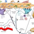
Patients with AD display skin-barrier impairment due to epidermal filaggrin deficiency caused by exogenous and/or inherited factors. These factors include a low relative humidity, excessive UV exposure, water, detergents, topical corticosteroids, and filaggrin gene ( FLG ) mutations. Epidermal filaggrin deficiency affects several important pathways, which puts further stress on the barrier.
The dysfunctional skin barrier makes patients with AD significantly more susceptible to irritant exposure and increases their risk of developing ICD. For example, application of an irritant sodium lauryl sulphate (SLS) (0.5% of SLS in an aqueous solution) result in a significantly higher skin reaction and transepidermal water loss in patients with AD irrespective of their FLG mutations status compared with controls.
Epidemiology of hand eczema in patients with atopic dermatitis
In 1985 Rystedt found an association between the occurrence of AD in childhood and presence of HE in adulthood. Since then, several studies have confirmed the potent association between these clinical entities, with the highest prevalence of HE in adolescents and young adults. Rystedt showed that HE is most common in patients with AD aged 25 to 29 years, likely because of a combination of risk factors including domestic and occupational wet work.
The association between AD and HE is strongest in patients with severe AD, in patients with AHE since childhood, and in patients with persistent AD on other parts of the body. Furthermore, AD is associated with more severe HE ; the resolution of HE is less likely in patients with AD compared with patients without AD. In general, studies that examined the association between AD and HE have not addressed the difference between AHE and HE due to ICD and/or ACD. Although it has been a topic of intensive discussions, it remains unclear whether AD also increases the risk of ACD; but it is likely nickel allergy and AD are associated. It is well known that FLG mutations represent a major predisposing factor for AD ; mutation carriers have characteristic palmar hyperlinearity, xerotic skin, as well as incidental skin fissures. Epidemiologic studies have examined whether FLG mutations increase the risk of HE, and it seems that mutations primarily increase the risk of HE in the context of AD. Affected patients have characteristic early onset of HE and tend to have more persistent and severe disease.
Most studies investigating the risk of occupational HE (OHE) in patients with AD have not addressed the difference between OHE due to ICD and ACD, respectively. However, Rystedt found that 77% of OHE cases were due to ICD, whereas only 6% were due to ACD. Studies have found that patients with AD have an increased risk of developing OHE when in health care, hairdressing, and metalwork. Furthermore, patients with AD tend to have more severe OHE, which results in a higher number of sick-leave episodes, job loss, and use of topical corticosteroids. Interestingly, a recent questionnaire study found the prevalence of AD to be significantly lower among hairdresser apprentices compared with general population controls. This finding indicates that patients with AD now may tend to avoid risk occupations, likely as a result of information given by physicians.
Related posts:
 Atopic Dermatitis: A Heterogeneous Disorder
 Public Health Burden and Epidemiology of Atopic Dermatitis
The Long-Term Course of Atopic Dermatitis
Long-Term Treatment of Atopic Dermatitis
Special Considerations for Therapy of Pediatric Atopic Dermatitis
Public Health Burden and Epidemiology of Atopic Dermatitis
The Long-Term Course of Atopic Dermatitis
Long-Term Treatment of Atopic Dermatitis
Special Considerations for Therapy of Pediatric Atopic Dermatitis
 Adjunctive Management of Itch in Atopic Dermatitis
Stay updated, free articles. Join our Telegram channel

Full access? Get Clinical Tree


