Lymphomatoid papulosis


Lymphomatoid papulosis


Rachel S. Klein, Elisha Singer, Jacqueline M. Junkins-Hopkins, Carmela C. Vittorio, Alain H. Rook and Ellen J. Kim


Evidence Levels:  A Double-blind study  B Clinical trial ≥ 20 subjects  C Clinical trial < 20 subjects  D Series ≥ 5 subjects  E Anecdotal case reports


image

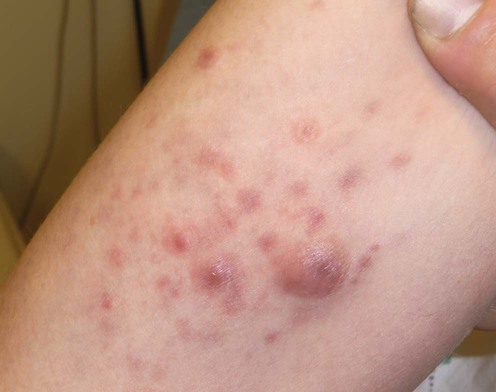
Lymphomatoid papulosis (LyP) is a distinct subset of CD30+ lymphoproliferative disorders in the World Health Organization/European Organization for the Research and Treatment of Cancer (WHO/EORTC) classification of cutaneous lymphomas, defined histologically by a variable infiltrate of CD30+ lymphocytes, in conjunction with the clinical presentation of recurrent, self-healing papulonodules. The lesions evolve into a crusted or necrotic stage, which often heal with a scar. Less common presentations include regional and oral disease. LyP affects adults and children as young as 11 months of age and may persist for years to decades. Approximately 5–25% of patients with LyP may present with or develop a lymphoproliferative malignancy, such as mycosis fungoides (MF – cutaneous T-cell lymphoma, CTCL), anaplastic large cell lymphoma (ALCL), and Hodgkin’s or non-Hodgkin’s lymphomas. There may also be a slight risk of developing a non-hematologic neoplasm. The same clone has been documented in patients with LyP and the associated lymphoma (including MF and ALCL), supporting the concept that LyP lies within the spectrum of CTCL. Clinical and histologic overlap may also be seen with LyP and pityriasis lichenoides et varioliformis acuta.


Management strategy


Most importantly, the management of LyP begins with understanding the natural history of the disease. LyP is defined as a recurrent, self-healing (remission of every individual lesion), papulonodular eruption, with histology suggesting CTCL. There are four histologic variants, including type A (wedge-shaped infiltrate containing eosinophils and histiocytes), type B (epidermotropism, resembles MF), type C (cohesive sheets of CD30+ cells, resembles ALCL), and type D (CD8+, resembles primary cutaneous aggressive epidermotropic CD8+ cytotoxic T-cell lymphoma). Clinical evaluation should include a complete history assessing for atypical features, including previous lymphoid neoplasms, constitutional symptoms, lymphadenopathy, or laboratory abnormalities on complete blood counts, blood chemistry, and lactate dehydrogenase. Patients with abnormal findings should undergo CT or PET/CT scanning to exclude a systemic lymphoma. Distinguishing LyP from primary cutaneous ALCL is important, although this may be difficult because of clinical and histologic overlap, including spontaneous regression.


Persistent lesions greater than 2–3 cm in diameter favor ALCL; however, some patients do not fit well into either category. Such borderline cases have similar biologic behavior to LyP and can be managed as such. Treatment of LyP should be tailored to the disease burden, because most modalities have not been proven to alter the natural course of LyP, nor do they prevent the development of extracutaneous lymphomas. Treatment should be reserved for symptomatic or cosmetically bothersome cases. First-line therapies include topical corticosteroids, low-dose methotrexate, and 8-methoxypsoralen with UVA (PUVA). A response may be seen within the first few weeks of treatment, including the development of fewer lesions, shortening of the lifecycle of individual lesions, and occasionally induction of remission. Large or borderline lesions can be treated with local radiotherapy or excision. Therapies that are beneficial in MF may also be used, including topical carmustine (BCNU), topical mechlorethamine (nitrogen mustard), and biologic agents such as interferon-α, interferon-γ, and the retinoid X receptor agonist bexarotene. Topical bexarotene, in a region of localized disease, may prevent recurrence of the condition. Similarly, topical imiquimod cream 5% can hasten resolution of lesions. In severe cases, the anti-CD30 antibody, brentuximab, may also be efficacious; however, this must be balanced by the risks of peripheral neuropathy, neutropenia, and rarely, progressive multifocal leukoencephalopathy (PML). Multiagent chemotherapy is not indicated, despite histologic features that may suggest ALCL. LyP can recur for decades, requiring careful consideration of treatment side effects and continued monitoring for the development of lymphoma.


Specific investigations


Primary and secondary cutaneous CD30+ lymphoproliferative disorders: a report from the Dutch Cutaneous Lymphoma Group on the long-term follow-up data of 219 patients and guidelines for diagnosis and treatment.

Bekkenk MW, Geelen FAMJ, van Voorst Vader PC, Heule F, Geerts ML, van Vloten WA, et al. Blood 2000; 95: 3653–61.


Guidelines are proposed for the diagnosis and treatment of patients with CD30+ lymphoproliferative disorders based on long-term follow-up of 219 patients, 118 of whom had LyP (4% had tumors). Fifty-two of 118 patients received no treatment or topical corticosteroids. The remainder received a variety of standard treatments, none of which were associated with complete, sustained remission. Staging of patients with type C LyP failed to reveal extracutaneous disease. Nineteen percent developed an associated lymphoma. Induction of remission of the secondary lymphoma had no effect on the natural course of LyP. The calculated risk for extracutaneous disease was 4% at 10 years.


CD30+ cutaneous lymphoproliferative disorders: the Stanford experience in lymphomatoid papulosis and primary cutaneous anaplastic large cell lymphoma.

Liu HL, Hoppe RT, Kohler S, Harvell JD, Reddy S, Kim YH. J Am Acad Dermatol 2003; 49: 1049–58.


In 19 of 31 cases with LyP, a higher than previously reported associated coexisting hematolymphoid malignancy (61% with one or more) was noted. Most had MF, which occurred before, during, or after the diagnosis of LyP. Some had two hematolymphoid malignancies. Three progressed to ALCL, with an interval ranging from 77 to 152 months. The overall 5- and 10-year survival of patients with LyP was 100% and 92%, respectively, and none died of LyP. Those with ALCL had a favorable course. LyP subtype did not predict the risk of associated malignancy.


The higher association with malignancy may represent selection bias.

Stay updated, free articles. Join our Telegram channel

Aug 7, 2016 | Posted by in Dermatology | Comments Off on Lymphomatoid papulosis

Full access? Get Clinical Tree

Get Clinical Tree app for offline access