Lymphocytoma cutis


Lymphocytoma cutis


Fiona J. Child and Sean J. Whittaker


Evidence Levels:  A Double-blind study  B Clinical trial ≥ 20 subjects  C Clinical trial < 20 subjects  D Series ≥ 5 subjects  E Anecdotal case reports


image


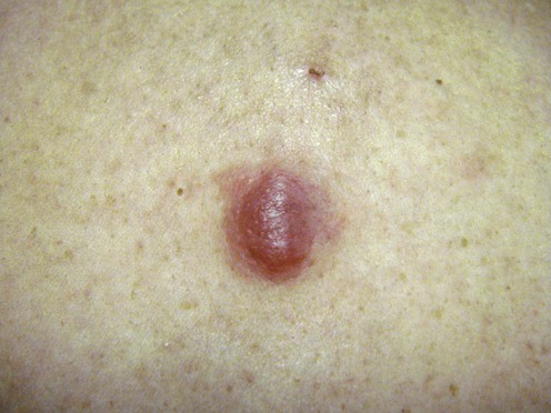
Lymphocytoma cutis (cutaneous lymphoid hyperplasia, cutaneous B-cell pseudolymphoma, Spiegler–Fendt sarcoid) is an entity encompassing a spectrum of benign B-cell lymphoproliferative diseases that share clinical and histopathologic features. Various stimuli can induce lymphocytoma cutis, but in most cases the cause is not known. It is more common in females, with a female-to-male ratio of 3 : 1. Most cases are characterized by localized erythematous, plum-colored nodules and plaques that may be difficult to distinguish from cutaneous B-cell lymphoma. Less frequently the generalized form may present with multiple miliary papules that measure a few millimeters in diameter. Lymphocytoma cutis secondary to Borrelia infection is most frequently seen at sites where skin temperature is low, such as the earlobes, nipples, nose, and scrotum.


Management strategy


A skin biopsy for histopathology and immunohistochemistry is required to confirm the diagnosis, but the distinction between lymphocytoma cutis and cutaneous B-cell lymphoma may be difficult on both clinical and histopathologic evaluation. There are no agreed histologic criteria; however, features that suggest lymphocytoma cutis include well-formed, non-expanded, reactive germinal centers, the majority of the infiltrate consisting of small round lymphocytes with a B:T cell ratio of <3 : 1 and polytypic expression of kappa and lambda light chains. A further feature is the presence of numerous tingible body macrophages within the lymphoid follicles. Molecular analysis of the immunoglobulin heavy chain gene has shown that a significant proportion harbor B-cell clones, which suggests that many cases previously thought to be lymphocytoma cutis represent indolent low-grade primary cutaneous B-cell lymphomas (PCBCL). Therefore, in cases with a detectable B-cell clone a careful evaluation to exclude systemic disease (a thorough clinical examination, thoracoabdominopelvic CT scan, and bone marrow biopsy) is required, with adequate long-term follow-up.


A history of possible stimuli known to cause lymphocytoma cutis should be sought; these include Borrelia burgdorferi infection, Leishmania infection, trauma, vaccinations, allergy hyposensitization injections, ingestion of drugs, arthropod bites, acupuncture, gold pierced earrings, tattoos, treatment with leeches (Hirudo medicinalis), and post herpes zoster scars, but the majority of cases are of unknown etiology.


The course of the disease varies but tends to be chronic and indolent, and some lesions may resolve spontaneously without treatment. There is no therapy of proven value for lymphocytoma cutis, with only anecdotal case reports and small series reported and no clinical trials in the literature.


If a cause can be identified, the causative agent should be removed. If infection with Borrelia burgdorferi is suspected, treatment with appropriate antibiotics (amoxycillin 500–1000 mg three times per day, or doxycycline 100 mg two to three times per day for at least 3 weeks) should be initiated.


Localized disease can be treated by simple excision and may respond to intralesional injection of corticosteroids, local irradiation, or intralesional interferon-α. More widespread (generalized) disease is traditionally treated with oral antimalarials, most commonly hydroxychloroquine (maximum dose 6.5 mg/kg/day); however, lesions may fail to respond to treatment or may recur following cessation of therapy. Other treatment modalities include subcutaneous interferon-α and oral thalidomide. Effective responses to destructive therapies, including cryotherapy and the argon laser, have been reported. A subtype of generalized lymphocytoma cutis may be exacerbated by light, and therefore sun avoidance and the use of sun block are important.


Specific investigations


Stay updated, free articles. Join our Telegram channel

Aug 7, 2016 | Posted by in Dermatology | Comments Off on Lymphocytoma cutis

Full access? Get Clinical Tree

Get Clinical Tree app for offline access