Fig. 22.1
Tension-free lymphatic–lymphatic end-to-end anastomosis technique. With permission from Baumeister RGH. Mikrochirurgie peripherer Lymphgefäße. In: Berger A, Hierner, R eds. Plastische Chirurgie: Band I Grundlagen Prinzipien Techniken. Springer © 2003, pp 213–217
Also because of the tininess of the vessels high magnifying microscopes have to be used. The finest instruments should be used and also the threads should be accustomed as much as possible to the needs of the lymphatic vessels. In the rat experiments it was found, that absorbable suture material showed less disturbance in the vicinity of the lymphatic anastomoses compared to non-absorbable material, where in relation to the vessels extended foreign body material has been observed [12].
Anastomoses between lymphatic vessels have a great advantage compared to anastomoses between lymphatic vessels and blood containing vessels. The coagulation abilities of lymphatic fluid are much lower compared to these of blood. Occlusion of anastomoses between lymphatic vessels is therefore rare.
Furthermore, lymphatic vessels showed in the experiments performed by Danese [13] the ability of building connection by themselves if lying next to each other. These effects may be one of the explanations about the high patency rate in lymphatico-lymphatic experimental anastomosing procedures. When anastomosing within the lymphatic transporting system there is no danger of an adverse pressure gradient which may be the fact by connecting lymphatic vessels to the peripheral venous system.
Patients should get the full range of conservative treatment consisting of manual lymphatic drainage and elastic stockings for at least half a year because during this time period also transient edemas are known. By that way he additionally got to know what is possible for him by the non-operative treatment. He may make up his mind whether this procedure fits for him or if it does not fulfill his expectations. He now is better willing to take a risk which is especially immanent to all invasive treatment options.
The most important factor for lymphatic vessel grafting is the possibility of harvesting lymphatic grafts. At least one lower extremity has to be edema free clinically. Additionally a normal lymphatic outflow prior to the harvest should be documented by a lymphatic scintigraphy.
The patient has to be ready for only a limited burden. This is a general anesthesia which lasts for about 2–4 h. The intervention itself takes place only within the subcutaneous tissue and a minimal blood loss has to be expected.
Harvesting of the Grafts
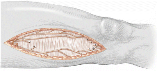
At the inner aspect of the thigh exists the ventromedial bundle. There up to 16 lymphatic collectors run in an almost parallel manner between the two narrowing areas of the lymphatic vascular system in the lower extremity: the knee region and the groin [14]. They are connected with side branches. This allows harvesting grafts with peripheral side branches giving the possibility to perform a greater number of peripheral lymphatic anastomoses than taking out lymphatic main collectors. Two to three collectors are mostly used (Fig. 22.2).


Fig. 22.2
Harvesting lymphatic vessels from the patient’s thigh. With permission from: Baumeister RGH. Lymphgefäßtransplantation an der oberen Extremität. In: Berger A, Hierner R, Eds. Plastische Chirurgie Extremitäten. Springer © 2009
In order to alleviate harvesting the grafts, patent blue™ is injected subdermally within the first and second web space. Within about 15 min lymphatic vessels at the thigh get stained. It is easy therefore to follow the vessels starting with an incision underneath the groin between the femoral artery and the greater saphenous vein. The incision ends proximal to the knee region. Care is taken to leave untouched stained lymphatic vessels behind in order to ensure a sufficient remaining lymphatic transport.
In order to reach ascending lymphatic vessels at the opposite leg the lymphatic grafts remain attached to the inguinal lymph nodes at the harvesting side. The peripheral endings of the grafts are occluded with a 6–0 thread with long endings. The grafts can be easily manipulated and pulled via an artificial tunnel above the symphysis to the opposite leg. The transected lymphatic collectors are sealed either by coagulation or by suturing to avoid lymphatic leakage.
For free transfers the lymphatic grafts are cut through beneath the inguinal lymph nodes and also occluded with a 6–0 thread providing long endings for further manipulation. In this case the peripheral openings of the grafts remain open. The grafts are freed from bigger fat lumps to reduce friction later on by gliding through artificial tunnels provided by silicone tubes.
Arm Edemas
Arm edemas are mostly caused by interference with the content of the axilla by surgery or irradiation, mostly in women suffering from mammary carcinomas. However, also treatment of male mammary carcinomas and lymph node resections in cases of Hodgkin disease are reasons for the development of edemas.
In these cases the axillary region has to be bypassed.
In front of the obstacle for the lymphatic transport an oblique incision is made above the vascular bundle. Ascending lymphatic main collectors run just above the fascia. The search starts by blunt meticulous dissection of the subcutaneous tissue. Small lymphatic vessels will be seen superficially. However, it should be looked for the greater vessels running more deeply. In contrast to small nerves, which are bright white and show oblique stripes, lymphatic vessels a grey looking and found between the fat tissue and also in the neighborhood of greater veins.
The next undisturbed lymphatic vessels are found at the neck. Below the sternocleoid muscle major lymphatic vessels transport lymph from the head to the venous angulation. The lymphatic vessels and the lymph nodes are imbedded in the fat tissues lateral to the internal jugular vein. They can be approached via a small oblique incision. An injection of patent blue™ subdermally behind the ear may facilitate to find the thin-walled lymphatic vessels. Alternatively lymphatic–lymph nodal anastomoses may be performed [15].
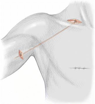
Between the incisions at the upper arm and the neck, a tunnel is created by blunt preparation using a long forceps. A silicone tube Charrier 18 with an appropriate length to exceed the length of the tunnel is prepared containing a long thread. The tube is inserted and the thread on the peripheral ending of the grafts is connected to the peripheral part of the thread within the tube. The graft is now pulled within tube until it appears at the neck. The graft is fixed manually at the neck and the tube is retracted towards the upper arm incision. Now the graft lies in place within the subcutaneous tissue. The proximal part is directed towards the neck, which means that the graft is situated in the appropriate direction to transport lymph from the peripheral ones towards the central anastomoses (Fig. 22.3).


Fig. 22.3
Bridging a lymphatic gap at the axilla with autologous lymphatic vessels: lympho-lymphatic anastomoses at the upper arm and the neck. With permission from: Baumeister RGH. Lymphgefäßtransplantation an der oberen Extremität. In: Berger A, Hierner R, Eds. Plastische Chirurgie Extremitäten. Springer © 2009
The anastomoses between the lymphatic vessels are performed mostly in an end-to-end fashion, sometimes also in an end-to-side version. Experimental data have shown that also anastomoses between lymphatic vessels and lymph nodes are possible and show also a high patency rate [16]. In this case the capsule is opened, giving access to the outer sinus close to the efferent lymphatic vessels. The anastomosis is performed with about four single stitches using smallest available suture material.
Stay updated, free articles. Join our Telegram channel

Full access? Get Clinical Tree


