Lower Abdominal Wall Reconstruction With Lateral Thigh-Based Flaps
Dhivya R. Srinivasa
Jeffrey H. Kozlow
DEFINITION
Abdominal wall reconstruction is required when partial- or fullthickness defects of the abdominal wall do not allow for primary fascial and/or skin closure. This situation can arise in a variety of clinical situations, sometimes presenting as an intraoperative consult requiring immediate closure. Defects should be evaluated for missing components of the abdominal wall including the skin, subcutaneous tissue, fascia, and musculature. Reconstruction should aim to close the intra-abdominal compartment while minimizing the risk of future hernia development. Techniques to reapproximate the midline fascia for a ventral hernia are reviewed in a separate chapter. Here, we will focus on restoring soft tissue where it is deficient in the lower abdomen.
ANATOMY
The anatomy of the abdominal wall is most readily described in layers. Most superficial is the skin and subcutaneous tissues. Within the subcutaneous tissues is the Scarpa fascia, a layer of variable thickness that provides some additional support to a repair. The critical component of the abdominal wall is the myofascial layer. This layer includes the paired, vertically oriented rectus abdominis muscles adjacent to the midline along with the internal oblique, external oblique, and transversalis muscles laterally. The investing fascia provides significant strength to the abdominal wall and includes both an anterior sheath and a posterior sheath. The central fascia around the rectus abdominis muscles extends from the oblique musculature. In the upper abdomen, the anterior and posterior sheaths are of similar strength; however below the arcuate line, the posterior sheath is much weaker as only the transversalis fascia contributes to the fascia deep to the rectus abdominis muscles. FIG 1 depicts the layers of the abdominal wall.
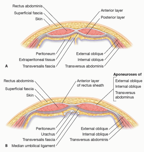 FIG 1 • Layers of the abdominal wall. A. Axial view of abdominal wall above the arcuate line. B. Axial view of abdominal wall below the arcuate line. |
Blood supply to the abdominal wall derives from multiple vessels with notable overlap in distribution. Centrally, the superior and inferior epigastric systems supply the abdominal wall. This is further enriched by branches from the internal mammary artery, specifically musculophrenic branches, as
well as the superficial epigastric vessels. Laterally, the circumflex vessels from the iliac artery (deep and superficial) add to the redundancy in blood supply. Innervation is via subcostal nerves (from T12) and thoracoabdominal nerves from T7-T11. These ventral primary rami branches travel between the internal oblique and transverse abdominal muscles. This is the target for the TAP (transverse abdominis plane) block that is used to anesthetize the abdominal wall in select procedures.
well as the superficial epigastric vessels. Laterally, the circumflex vessels from the iliac artery (deep and superficial) add to the redundancy in blood supply. Innervation is via subcostal nerves (from T12) and thoracoabdominal nerves from T7-T11. These ventral primary rami branches travel between the internal oblique and transverse abdominal muscles. This is the target for the TAP (transverse abdominis plane) block that is used to anesthetize the abdominal wall in select procedures.
A thorough understanding of the various meshes used in abdominal wall reconstruction (including the various synthetic and biologic materials) is critical for surgeons planning to use these materials. However, details on this topic are beyond the scope of this chapter.
PATHOGENESIS
Abdominal wall defects can arise from a variety of scenarios: trauma, cancer, and hernia reconstruction being the most common. Acquired hernias present as a fascial defect and often require abdominal wall myofascial advancement flaps and/or mesh-related techniques. This chapter will focus on closure of soft tissue defects that arise from traumatic injury and oncologic resections. However, some of the principles used for acquired hernia repair, such as component separation, can also play a role in traumatic and oncologic reconstructions. In trauma, all devitalized tissue should be debrided prior to closure. With respect to cancer, margins should be clear and the need for adjuvant radiation should be consider when choosing a reconstructive technique.
PATIENT HISTORY AND PHYSICAL FINDINGS
In addition to the usual key components of a patient’s history, the following specifics should also be documented:
Prior abdominal surgery
Include laparoscopic operations and cesarean sections
Need for stoma postresection. If an ostomy is planned, the estimated location for the stoma should also be noted.
History of radiation therapy or planned radiation therapy
Medical comorbidities that will affect wound healing including diabetes mellitus, steroid use, tobacco use, or connective tissue disorders
Prothrombotic disease states should be noted and a Caprini score should be calculated. Appropriate venous thromboembolism prophylaxis should be tailored accordingly.1
Each patient should be examined thoroughly and all prior incisions should be noted.
Evaluation of the thigh for adequate tissue to support transfer of the necessary tissue from this area to the abdomen.
Incisions within the estimated location of the donor flap should be noted. We also suggest an evaluation of body habitus in this region as thick or thin flaps may be harvested depending on the reconstructive needs.
IMAGING
No specific imaging is necessary for the reconstructive portion of the case. However, most patients will have an abdominal computed tomography (CT) scan, which can be used for planning purposes to understand the anticipated defect. In oncologic cases, specifically note involvement of the skin, anterior sheath, musculature, posterior sheath, and intraperitoneal or retroperitoneal contents. CT angiography of the thigh can be performed in select cases to evaluate perforator location or regional anatomy in complex situations.
SURGICAL MANAGEMENT
Preoperative Planning
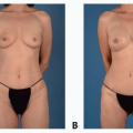
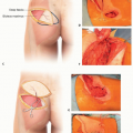
Preoperative planning is incumbent on anticipation of the size and depth of the defect. Whether full-thickness abdominal wall resection is necessary will dictate if fascial advancement and/or mesh will be necessary. The location of the defect is also important to note, as it will affect potential flap options. FIG 2 illustrates various abdominal wall defects and how they can be covered with a thigh-based flap.
Inguinal defects can often be addressed with ipsilateral tensor fascia lata (TFL) based flaps. These flaps provide strong fascial tissue if needed for reconstruction of the inguinal ligament.
Lower abdominal (below the umbilicus) and flank defects can be addressed with flaps based off the descending branch of the lateral femoral circumflex system such as the anterolateral thigh (ALT) flap, vastus lateralis, or rectus femoris flaps.2,3
Midabdominal defects can be addressed with component separation techniques similar to ventral hernia repairs owing to the freedom of the abdominal wall tissue away from the fixed bony pelvis and rib cage.
Upper abdominal defects are the most difficult as thighbased flaps will typically not transpose to the upper abdomen without vein grafts.
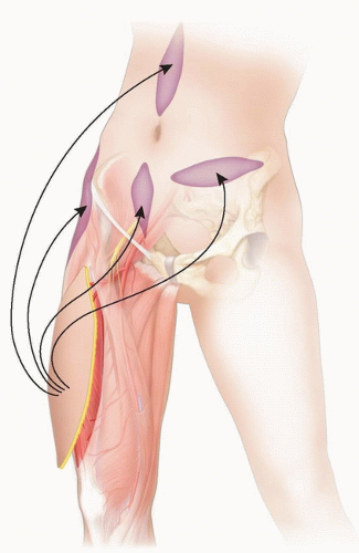
FIG 2 • Lateral thigh-based flaps are a versatile option for a variety of lower abdominal wall defects.
Preoperative discussion with the patient should include possible donor sites, skin grafting, and potential use of mesh.
Any planned stomas or existing stomas should be discussed so modifications can be made accordingly. Existing stomas likely pass through the rectus abdominis muscle. Future stomas should be marked preoperatively.
In cases where primary myofascial repair may be possible, preoperative administration of Botox can be helpful if administered in advance. Typically, up to 100 units per side can be given at least 1 to 2 weeks prior to surgery to optimize the timing of neuromuscular blockade. The goal is to allow maximal relaxation of abdominal wall musculature in anticipation of myofascial advancement for closure.4
Positioning
The patient will likely be positioned supine for the abdominal portion of the surgery. All of the lateral thigh-based flaps are most easily harvested in the supine position as well.
A safety belt should be used for all cases, but this most often is placed around the chest area.
A warming blanket should be placed to optimize normothermic core temperatures during the case.
SCDs should be placed bilaterally as the critical areas of the thigh need to be prepped only to the knee and do not require intraoperative rotation of the leg.
The thigh should be prepped circumferentially. The contralateral thigh should also be available in the event skin graft is needed for closure of the defect or donor site.
Approach
Preoperative markings should be made in the preoperative area, prior to patient positioning in the OR.
Anatomic Landmarks: The anterior superior iliac spine (ASIS) and the superolateral patella are identified. A line connecting these two points is drawn. For an ALT flap, the midpoint of this line will be near the central axis of the skin paddle. FIG 3 depicts relevant anatomic landmarks and the axis of the skin paddle.
If a TFL flap is planned, the incision can be adjusted superiorly (cephalad).
If a rectus femoris muscle flap is planned, the incision can be adjusted medially.
If an ALT flap is planned, there are two main approaches to identifying perforators and designing the skin paddle:
Some surgeons define the “A, B, and C” perforators. The perforators are most commonly found approximately 1.5 cm posterior to the line between the ASIS and superior lateral patella. The center point of this line should be marked as perforator “B.” Perforators “A” and “C” are 5 cm proximal and distal to “B.”5
Another approach is to draw a 3-cm-radius circle around the midpoint of the axis line, defining the most common location of skin perforators. A Doppler can be used to confirm and mark the location of these perforators. The skin paddle can be centered over these perforators. Most often the perforators are found in the inferolateral quadrant of the circle (FIG 4).Related posts:

Stay updated, free articles. Join our Telegram channel

Full access? Get Clinical Tree