Fig. 46.1
Reconstruction of the abdominal wall using the PANO protocol. The rectus abdominis muscles can be seen along the vertical axis of the abdominal wall from the xiphoid down to the pubis. The dark image between the upper and lower abdomen is the umbilical region
46.4 Statistical Analysis
F-test was used to compare the variance between both groups. Student t-test was used to compare the age average between groups. Sturges’ test was used to evaluate the characteristics of the groups. Z-test was used to evaluate the efficacy of plication. Shapiro’s test was used to evaluate the distribution of diastasis measurements. The statistical tests were performed at the significance level of 0.05.
46.5 Results
Patients in group A were compared to those from group B regarding age, BMI (Table 46.1), number of smokers, physical activity, and number of pregnancies using Student t-test, F-test, and Sturges’ test (Table 46.2). Sturges’ test was used for comparisons regarding class variance. Student t-test was used for comparisons regarding age variance. Statistical significant difference for p ≤ 0.05.
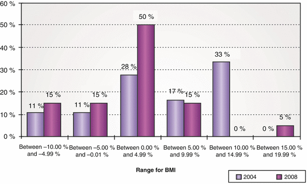
Table 46.1
Evolution of BMI – 2004 to 2008
Table 46.2
Comparison between groups regarding age, BMI, number of smokers, physical activity, and number of pregnancies
Characteristics | Group A (5 years – PO) | Group B (1 year – PO) |
|---|---|---|
Age | 34–67 p = 48 | 23–68 p = 43 |
BMI at the time of surgery | 20.96–27.06 p = 23.73 | 21.62–29.49 p = 24.72 |
Number of smokers | 4 (22.22 %) | 3 (15 %) |
Number of patients with regular physical activity | 10 (56 %) | 11 (55 %) |
Number of previous pregnancies | 18 (100 %) | 19 (95 %) |
In group A, BMI ranged from 20.96 to 27.06 kg/m2 at the moment of the postoperative ultrasound. In this group, four patients had lost weight and 14 patients increased weight after the surgery. In group B, these numbers varied from 21.62 to 29.49 kg/m2, with two patients showing no weight variation, six patients having weight loss, and 12 patients having gained weight (Fig. 46.1).
Five small hernias were found intraoperatively and were promptly corrected: one patient from group A (6 %) and four from group B (20 %). These hernias had not been detected by the preoperative ultrasound evaluation. Surgical correction of these hernias was done by the simple approximation of the aponeurotic edges.
As regards complications, only one patient in group A had a significant hematoma within the first 24 h postoperative period, requiring a surgical revision. Two other patients from group B presented with serosanguinous collection, which was treated with serial aspirations. No other complications were noted.
Clinical examination revealed, in one patient from group B, a slight diastasis of the upper abdomen. All other patients denied any complaints of pain or discomfort along the midline while performing exercises, suggesting that there was only a single case of clinical recurrence of muscle diastasis.
In group A (5 years postoperative), all measurements of the distance between the aponeurotic edges were equal to zero after the surgery (Fig. 46.2). Two patients in group B (1 year postoperative) presented recurrence of the diastasis, in which an increase in the distance between the aponeurotic edges in the upper abdomen was noted (Fig. 46.3). This distance was 0.87 cm in one patient and 1.30 cm in the other patient. In both of them, diastasis recurred in the vicinity of the xiphoid. It was also noted with the US examination that there was a lateral insertion of the recti muscles on the costal margins. In these patients, there was a minimal bulging along the midline. However, these two patients presented no clinical complaint regarding this finding.
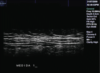
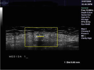
Fig. 46.2
There is no recurrence of rectus diastasis in this patient. Note that the recti muscles are close together in the midline after 5 years after the plication of the anterior rectus sheath
Fig. 46.3
In detail, the US examination reveals a distance of 0.87 cm between the aponeurotic edges in one of the two patients in group B that presented recurrence of diastasis
46.6 Discussion
Correction of the separation of the recti muscles has been performed since the 1960s [7, 9–14] with plication of the anterior rectus sheath to correct rectus diastasis being the most frequently performed procedure in this layer. Pitanguy described his approach, where the rectus sheath is maintained intact, and suturing of the borders is done, starting at the xiphoid down to the pubis, using nonabsorbable 2–0 nylon with inverted X knots.
Related posts:
 Abdominoplasty Combined with Cesarean Section: Discussion of the Evidence
Retrospective Analysis of Never Events in Panniculectomy and Abdominoplasty Patients and Their Financial Implications
Abdominoplasty Combined with Cesarean Section: Discussion of the Evidence
Retrospective Analysis of Never Events in Panniculectomy and Abdominoplasty Patients and Their Financial Implications
 No Drain Abdominoplasty with Progressive Tension Suture: The Logic, Simplicity, and Aesthetics
 Evolution of the High-Superior-Tension Lipoabdominoplasty: The High-Tension Abdominoplasty
 Circumferential Abdominoplasty
 Abdominal Wall Repair Post Hernia in Kidney and Liver Transplantation
Stay updated, free articles. Join our Telegram channel

Full access? Get Clinical Tree


