Livedoid vasculopathy


Livedoid vasculopathy


Bethany R. Hairston and Mark D.P. Davis


Evidence Levels:  A Double-blind study  B Clinical trial ≥ 20 subjects  C Clinical trial < 20 subjects  D Series ≥ 5 subjects  E Anecdotal case reports


image
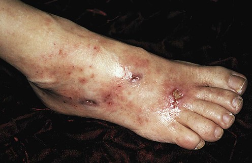
(From Lebwohl, M.G., 2003. The Skin and Systemic Disease: A Color Atlas, second ed. Churchill Livingstone, with permission of Elsevier.)


Livedoid vasculopathy, or livedoid vasculitis, is a painful ulcerative condition of the lower extremities with characteristic clinical and histopathologic features. The lesions of atrophie blanche, a term once synonymous with the disease, are typically present on the lower extremities and are characterized by smooth, porcelain-white lesions surrounded by punctate telangiectasia and hyperpigmentation. Central, shallow ulceration is often present. This condition is difficult to treat and often recalcitrant to therapy.


Management strategy


Appropriate diagnosis of livedoid vasculopathy is necessary before treatment options can be considered. Histologic identification of the characteristic segmental hyalinized appearance of the dermal blood vessels is important to exclude other causes of lower extremity ulcerative disease. Increasing numbers of reports suggest that the disease has a procoagulant predisposition, with both hereditary and acquired hypercoagulable states.


Typically shallow and numerous, the ulcerations in livedoid vasculopathy are painful and slow healing. Wound care is an important facet of treatment. Excellent dressings and topical products are available to treat chronic ulcerative diseases, and selection depends on the moisture content of the wound and the possibility of superinfection. Topical and oral antibiotics may be beneficial, and wounds should be cultured to determine appropriate sensitivity to medications. Pain management is also essential.


Because of the potential procoagulant mechanisms involved in disease etiology, medical therapy has traditionally centered on prevention and treatment of dermal vessel thrombosis and improvement of vascular perfusion. Medical management has included aspirin (acetylsalicylic acid), niacin (nicotinic acid), pentoxifylline, dipyridamole, warfarin, and danazol. Systemic corticosteroids are not considered a primary therapy; however, some patients’ conditions have improved with immunosuppressants in combination therapy. Psoralen with UVA (PUVA) has also been used. Patients with livedoid vasculitis recalcitrant to traditional management have been treated with minidose heparin, subcutaneous low-molecular-weight heparin injections, intravenous iloprost (a prostacyclin analog), intravenous immunoglobulin (IVIG), and a tissue-type plasminogen activator (tPA).


Specific investigations


Stay updated, free articles. Join our Telegram channel

Aug 7, 2016 | Posted by in Dermatology | Comments Off on Livedoid vasculopathy

Full access? Get Clinical Tree

Get Clinical Tree app for offline access