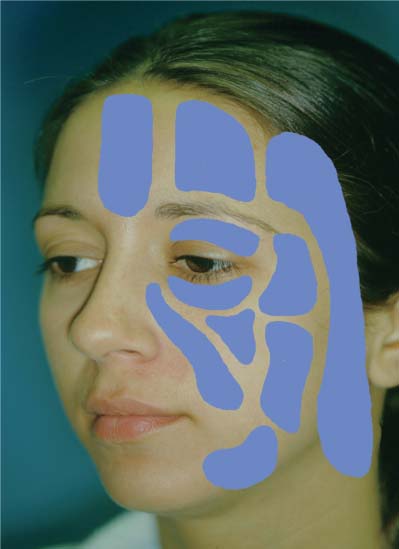
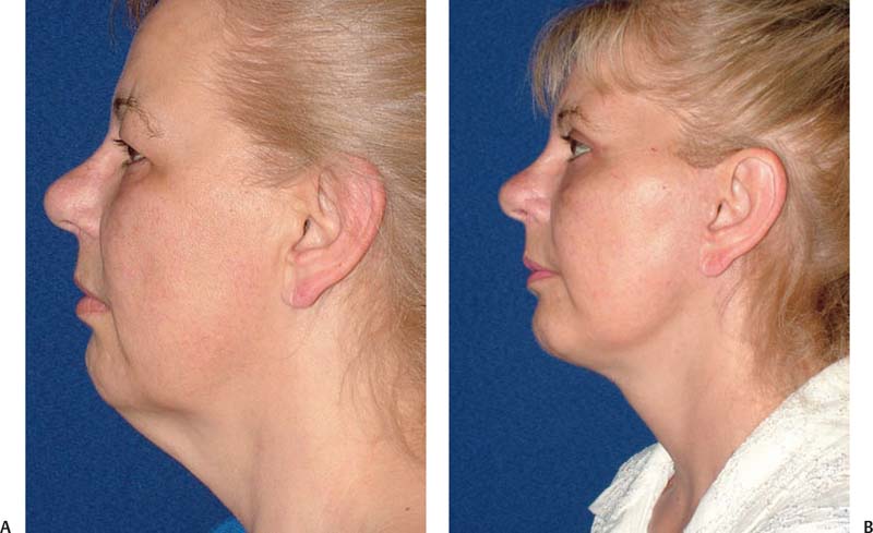
7 In the time between the present and its original description in 1976 by Dr. Giorgio Fischer,1 an Italian surgeon trained in otolaryngology, liposuction has become one of the most commonly performed cosmetic surgical operations in the United States. In 1978, two French physicians, Illouz and Fournier, vigorously embraced liposuction and further refined the procedure for it.2 Illouz introduced the “wet technique,” which utilized hyaluronic acid in hypotonic solution for injection before the performance of liposuction. Fournier first described syringe liposuction and the “criss-cross” technique, and was a noted and energetic international teacher of the procedure. Martin, an otolaryngologist in Los Angeles,2 first performed liposuction in the United States in 1982. Although quickly gaining popularity in the United States, the technique received some negative publicity when several patients experienced excessive bleeding and anesthetic complications postoperatively. In 1987, responding to these concerns, Dr. Jeffrey A. Klein, a California dermatologist, reported his 2-year experience with a “tumescent technique” for liposuction that was performed exclusively under local anesthesia.3 Before this time, the pain of liposuction required general or IV sedation. The tumescent technique revolutionized liposuction by eliminating the need for general anesthesia and by minimizing blood loss during the procedure. In 2000, Klein wrote a definitive book about tumescent liposuction entitled The Tumescent Technique,2 which covered in detail the major topics relevant to this procedure, including tumescent anesthesia, microcannular liposuction, and local anesthesia, and the pathophysiology, complications, pharmacology, pharmacokinetics, surgical technique, and postoperative care involved in the tumescent technique, as well as explanations of the special considerations for liposuction in each area of the body.2 More recent innovations in liposuction include power-assisted liposuction (PAL), ultrasonically assisted liposuction (UAL), and laser-assisted liposuction. Liposuction of the face and neck is a uniquely nuanced procedure. Unlike liposuction of the abdomen, thighs, or torso, which relies on the reduction of fat to restore aesthetic contour, the restoration of a youthful facial appearance in the aging patient is accomplished both by volume reduction and volume augmentation. It is therefore critical to understand the different fat compartments of the face and how they typically change with aging.4–6 The young face has a full look with a homogeneous distribution of both superficial and deep fat. The facial fat compartments transition seamlessly from one to the next, creating facial contours consisting of smooth arcs. In contrast, the aging face is characterized by a “hill-and-valley” appearance. There is an obvious demarcation between facial fat compartments, and the contours of the aging face are broken and irregular. These stigmata of aging result from the atrophy of certain fat compartments during the aging process, while other such compartments hypertrophy or remain unchanged. Facial fat atrophies in the temporal, buccal, periorbital, perioral, and forehead regions, and hypertrophies in the infraorbital and submental areas, jowl, lateral nasolabial folds, lateral labiomental creases, and lateral malar areas. Interestingly, the infraorbital area may show concavities and atrophic changes (the tear-trough deformity) with aging, or may develop bulging of infraorbital fat and festooning. Further variations in the distribution of facial fat depend on whether a patient has a heavy or a lean face. Thus, proper facial rejuvenation relies on appropriate rebalancing of the facial fat compartments. Hypertrophic areas may require fat removal and atrophic areas may need some form of volume augmentation. Although this may seem self-evident, it is not uncommon to see premature aging of the face caused by the over-reduction of a “hill” to match the contour of a “valley,” as is seen with hollowing of the eye when suborbital fat is overreduced in an attempt to reduce a tear-trough deformity. Conservative blending and balancing of tissue contours in the face should be foremost in the mind of the cervicofacial plastic surgeon. Liposuction of the face is done in the subcutaneous plane, in which the facial fat is divided into 10 independent compartments (Fig. 7.1). These include the nasolabial fold and malar (medial, middle, and lateral temporal [cheek portion]), forehead (central, middle, lateral temporal [forehead portion]), jowl, and orbital (superior, inferior, lateral) compartments. The nasolabial fat compartment lies anteromedially to the medial malar fat compartment and overlaps the jowl fat. The superior border of the nasolabial fat compartment is defined by the retaining ligament of the orbicularis oculi muscle. The malar fat compartment is just lateral to the nasolabial fat compartment and is superficial to the parotid gland. It is divided into distinct medial, middle, and lateral temporal compartmens and is bounded superiorly by the inferior and lateral orbital compartments and inferiorly by the jowl compartment. The lateral temporal compartment extends superiorly into the forehead region and inferiorly into the cervical region. Fig. 7.1 Rendering of the facial compartments of subcutaneous fat. The forehead fat compartment also comprises three distinct compartments. Of these, the lateral temporal compartment was mentioned above. The two other subcutaneous compartments of forehead fat are the central and middle compartments. The central compartment of forehead fat is located in the midline region of the forehead and extends inferiorly to the root of the nose. On either side of this compartment is the middle forehead fat compartment, which is bounded laterally by the lateral temporal compartment and inferiorly by the superior orbital compartment. The orbital fat compartment is composed of three unique subcutaneous compartments around the eye, consisting of the upper, lower, and lateral compartments. The upper and lower sub-cutaneous fat compartments are just adjacent to their respective eyelid tarsi. The lateral fat compartment is bounded superiorly by the inferior temporal septum and inferiorly by the septum of the superior cheek-fat compartment. The jowl-fat compartment is bounded medially by the depressor anguli oris muscle, to which it is adherent, and inferiorly by a membranous fusion of the platysma muscle. Besides the compartments described above, one other facial fat compartment can have significant impact on the shape and contour of the face: the buccal fat pad. Unlike the fat compartments described above, the buccal fat pad is a large, complex compartment that lies deep to the muscles of mastication. It is distinct from the subcutaneous jowl fat and lies deep to the subcutaneous musculoaponeurotic system (SMAS). In certain patients, buccal fat contributes to fullness of the lower cheek area. If prominent, buccal fat can be reduced by direct excision either through an intraoral approach or through an open approach during rhytidectomy, but is not accessible to reduction by liposuction. In contrast to the many distinct fat compartments of the facial region, the subcutaneous fat of the neck is continuous across the neck, although it is typically most prominent in the submental area beneath the chin. In nonobese individuals, submental fat may or may not be substantial. Significant submental fat in a thin patient is usually a heritable trait and not the result of aging. Liposuction in the neck is exceptionally safe provided it is limited to the supraplatysmal tissue layer. The operator working above the platysma is protected against injuring the facial nerve and major facial vessels. The bilateral platysma muscle, which extends into the face in the form of the SMAS, has its bony origin on the clavicle. From here it traverses the anterior neck and ascends over the mandible onto the cheek, where it is continuous with the SMAS of the lateral cheek. The platysma forms a critical barrier: superficial to it is the subcutaneous fat of the neck and lower cheek, as noted above. The profile of the neck is best appreciated from the lateral view, and the most important cephalometric parameter describing the youthful neck is the angle formed by the intersection of the anterior border of the neck and the submental plane.5,6 As the neck ages, gravitational changes and increased adiposity conspire to increase the cervicomental angle (CMA) and to obliterate the aesthetic features of a youthful neck. With accumulation of fat and increased laxity of the SMAS and skin in the submental area, the apparent intersection point of the border of the neck and submental plane (the cervical point) descends to a lower position in the neck, causing the intersection of these two elements to look less angulated (Fig. 7.2). In patients with prominent submental fat, liposuction of the neck is the single most effective surgical intervention for improving the angularity of the cervical point. Liposuction decreases fat volume and stimulates skin contraction, both of which raise the apparent cervical point.7,8 Several other anatomical relationships also determine the overall appearance and proportions of the neck. The subplatysmal fat lies immediately beneath the anterior aspect of decussated fibers of the conjoined platysma muscles and just caudal to the submental crease. Removal of some of this fat may reduce the perceived bulk of a full neck. However, the fat in this area is typically quite vascular and care must be taken when operating in this part of the neck. The hyoid bone is a critical structure in determining the proportions of the neck because it is the anchor point for the deep muscles of the anterior neck; its position affects the perceived fullness of the neck. An anteriorly located hyoid bone will increase fullness of the neck and decrease the perceived angularity between the jawline and neck. This is an important finding for the surgeon because it portends limitations to any rejuvenation procedure attempted in this region. The thyroid cartilage is another structure that can add perceived fullness to the neck. Liposuction must be performed carefully around this structure so as to not over-reduce the subcutaneous fat that overlies it. Such an error might make this cartilage more apparent, which would be particularly unacceptable in women. Fig. 7.2 (A) Poor CMA secondary to platysmal ptosis and excess submental fat. (B) Marked improvement of the CMA after platysmapexy and liposuction. Ancillary measures, such as platysmal plication (platysmaplasty) and cervicofacial rhytidectomy (facelift), also raise the cervical point.9 Whether used alone or in combination, both procedures can significantly enhance the neckline. Liposuction is defined as the removal of fat from deposits beneath the skin through a hollow cannula with the assistance of a powerful vacuum. Initially, liposuction was performed under general anesthesia using what was called the “dry technique” because it did not involve the injection of a local anesthetic agent into the fat before liposuction. There was no hemostasis within the adipose layer, and as a result, complications such as bleeding, the need for autologous blood transfusion, hematoma, irregularities in the skin, large-volume fluid shifts, and a prolonged recovery time were common. Remarkably, blood constituted ~30% of the tissue that was removed by liposuction with the dry technique. In an attempt to decrease such complications, surgeons began experimenting with different anesthetic solutions for infiltrating fat in preparation for liposuction by varying the concentrations of lidocaine and epinephrine in these solutions. With the progression from the “wet technique” to the “super-wet technique” and then to the “tumescent technique,” surgeons infiltrated more dilute anesthetic solutions in higher volumes into a given target area.3,10,11 The wet technique utilized commonly available commercial preparations of local anesthetic agents containing epinephrine, such as 1% lidocaine with a 1:100,000 dilution of epinephrine. Although the wet technique caused less blood loss than did the dry technique, blood loss was still dangerously excessive, with blood constituting ~15% to ~20% of the contents removed by liposuction. The super-wet technique utilizes a dilute local anesthestic agent in a volume less than half of that used for the tumescent technique (~0.5% lidocaine, 1:250,000 epinephrine). Surgical blood loss with the super-wet technique is greater than with the tumescent technique but significantly less than with the wet technique, with blood constituting only 8% of the fluid removed. Current methods of liposuction done with tumescent anesthesia minimize blood loss, reducing it to ~1 to 2% of the fluid removed, and eliminate the need for parenteral anesthesia. In one prospective randomized study of 112 patients, 9.7 mL of whole blood (~1%) was suctioned for each 1000 mL of fat removed. All patients were treated as out-patients, and there were no hospitalizations, transfusions, seromas, or other complications.12 The word “tumescent” means swollen and firm. The injection of a large volume of very dilute lidocaine (a local anesthetic agent) and epinephrine (a capillary constrictor) into subcutaneous fat renders the targeted fatty tissue swollen and firm, or tumescent. In the process, the following three critical goals are achieved: (1) the method of lidocaine administration blankets large areas of subcutaneous fat and consequently allows liposuction to be performed solely under local anesthesia; (2) the epinephrine component of the injectate constricts capillaries and sharply limits surgical blood loss; and (3) because of the fluid volumes injected into the subcutaneous region, no IV fluids are needed. An additional benefit of the tumescent technique is that the intense vasoconstriction it produces delays absorption of the lidocaine in the anesthetic solution. The local anesthetic remains trapped in the subcutaneous fat, providing 24 to 36 hours of significant postoperative analgesia that in turn decreases the requirements for postoperative use of narcotic analgesic agents. Other benefits of the tumescent technique include: (1) a hydrodissecting effect that allows easier and more uniform removal of fat; (2) elimination of the risks of general anesthesia; and (3) improved cost savings with the elimination of hospital anesthetia fees and facility fees. The tumescent technique is unquestionably the safest technique for liposuction. There has never been a reported death associated with tumescent liposuction done solely under local anesthesia.13,14 Even when general anesthesia is combined with the tumescent technique, liposuction is quite safe provided the volume of fat removed and the number of areas treated during a single surgery are not excessive. The standard Klein formula for the solution used in tumescent liposuction includes a mixture of saline (or lactated Ringer’s) solution, lidocaine, epinephrine, and sodium bicarbonate. Some surgeons choose to add triamcinolone to the solution. The solution may be delivered to the patient at room temperature, but warming of the solution provides greater patient comfort. Depending upon the clinical requirements, a tumescent anesthetic solution may contain a 5- to 40-fold dilution of the lidocaine present in commercially available formulations for local anesthesia. These formulations, used by dentists and anesthesiologists, typically contain 1 g of lidocaine and 1 mg of epinephrine per 50 mL of saline. In contrast, the tumescent solutions used for local anesthesia in liposuction contain ~1 g of lidocaine and 1 mg of epinephrine in 1000 mL of saline, representing an ~20-fold greater dilution when compared to commercially available formulations. The guidelines of the American Academy of Dermatology for care in liposuction15 recommended a maximum dosage of lidocaine of from 45 mg/kg to 55 mg/kg for liposuction under tumescent anesthesia. This is a relatively large dosage as compared with the 7 mg/kg that is widely accepted as the “safe maximum dose” for lidocaine with epinephrine used by anesthesiologists.16 How is it that tumescent liposuction under local anesthesia alone is safe despite the use of unprecedentedly large doses of lidocaine and epinephrine? The explanation for this remarkable safety is the extreme dilution of the tumescent local anesthetic solution. Large volumes of dilute epinephrine produce intense constriction of capillaries in the targeted fat, delaying the absorption of lidocaine and extending it across the 24 to 36 hours following infusion.17,18 Undiluted lidocaine and epinephrine are absorbed into the bloodstream in less than an hour. The dilution of these agents used in the tumescent technique reduces their potential toxicity in the following two ways: (1) a significant amount of the lidocaine/epinephrine solution is trapped in the fat cells and removed during the process of liposuction; and (2) because their absorption is greatly slowed, the peak blood concentrations of lidocaine and epinephrine present at any one time are greatly reduced.2,11,17,19 The anesthetic solution used in the tumescent technique can vary in composition. Typically, the contentration of the lidocaine component constitutes between 0.1 and 0.2% of the solution by weight, and the epinephrine concentration can range from 1: 500,000 to 1:1,000,000. The proportions preferred by the author are shown in Table 7.1. Tumescent liposuction in the head and neck is a particularly safe procedure when performed properly. Besides the advantages listed above, including the avoidance of systemic anesthesia, tumescent liposuction in the neck and jowls involves the infusion of less anesthetic solution (less than 500 mL) and the extraction of a far smaller volume of fat than do “whole-body” procedures. Nonetheless, use of the tumescent technique in the head and neck region does involve the use of larger volumes of anesthetic solution than do the wet and super-wet techniques, and creates appreciably more soft-tissue distortion. In inexperienced hands, asymmetric infusion of a tumescent anesthetic solution, loss of normal topographical landmarks, or both can lead to the asymmetric extraction of fat and suboptimal contouring of the jowl and neck. Thus the surgeon, understanding safe dosing parameters, may choose to adjust the concentration and volume of the infused anesthetic solution according to the surgeon’s practice preference.
Liposculpture of the Head and Neck
Understanding the Distribution of Fat in the Aging Face and Neck
Facial Anatomy
Fat Compartments

Topographic Landmarks

The Anesthetic Evolution of Modern Liposuction
Wet Technique
Super-wet Technique
Tumescent Technique
Tumescent Solution
Composition of Tumescent Fluid for Liposuction Containing Stay updated, free articles. Join our Telegram channel
Full access? Get Clinical Tree
 Get Clinical Tree app for offline access

|


