9
Lichen Planus and Lichenoid Dermatoses
Lichen Planus
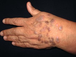
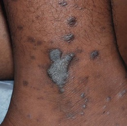
• Flat-topped (lichenoid) papules that are often polygonal in shape and purple in color may coalesce into plaques (Fig. 9.1); lesions usually resolve with hyperpigmentation (Fig. 9.2).
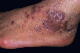
Fig. 9.1 Lichen planus. Violaceous papules and plaques with white scale and Wickham’s striae. Courtesy, Tetsuo Shiohara, MD.
• A characteristic finding is Wickham’s striae, a network of fine white lines on the surface of papules and plaques (Fig. 9.3).
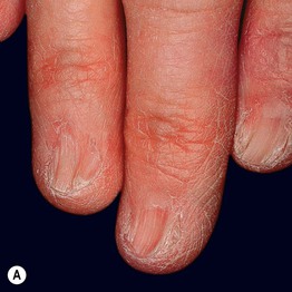
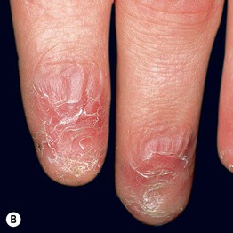
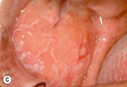
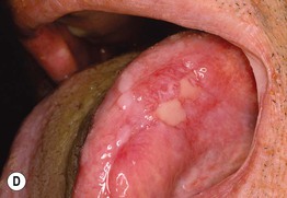
• There are multiple variants of lichen planus, from exanthematous to hypertrophic (Table 9.1; Figs. 9.4 and 9.5).
Table 9.1
Variants of lichen planus.
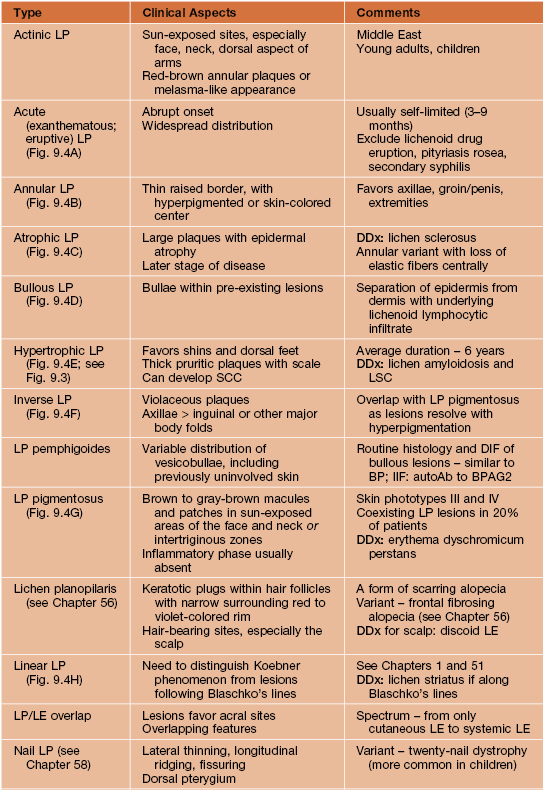

* If erosive variant plus lichenoid cutaneous lesions, DDx includes paraneoplastic pemphigus.
AutoAb, autoantibody; BP, bullous pemphigoid; BPAG2, bullous pemphigoid antigen 2 (type XVII collagen); DIF, direct immunofluorescence; HCV, hepatitis C virus; IIF, indirect immunofluorescence; LE, lupus erythematosus; LSC, lichen simplex chronicus; SCC, squamous cell carcinoma.
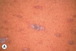
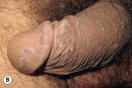
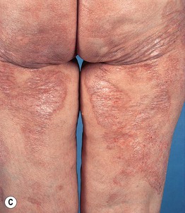
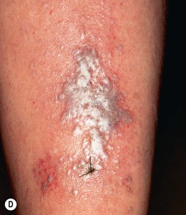
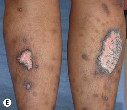
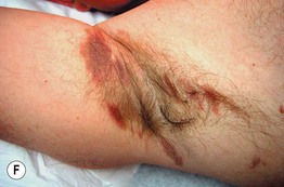
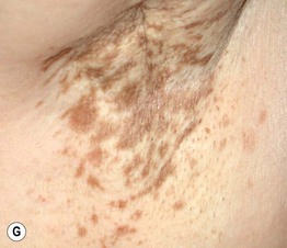
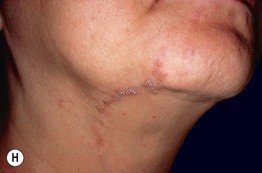
Fig. 9.4 Variants of lichen planus. A Exanthematous variant with multiple papulosquamous lesions. B Annular variant with thin elevated rim and central hyperpigmentation. C Atrophic variant with large, long-standing lesions. D Bullous variant with vesicobullae arising in pre-existing plaque. E Hypertrophic variant with thick plaques on the shins, a common location for this variant. F Inverse variant of the axillae; note the purple color. G Pigmentosus variant with only hyperpigmented lesions in an intertriginous zone. H Linear variant along Blaschko’s lines with follicular spines. B, Courtesy, Frank Samarin, MD; C, Courtesy, Tetsuo Shiohara, MD. F, Courtesy, Jeffrey Callen, MD.

