With the growth of new technology and products over the last 10 years, there has been an increased ability to improve a patient’s appearance with procedures that can be performed in an office setting, including laser procedures. Demand for these procedures has grown among all ethnic groups. Patients with ethnic skin can have varying response to lasers. This factor should be considered when planning their treatment. After laser treatment, Patients with ethnic skin are at greater risk for laser energy absorption by melanin, postinflammatory hyperpigmentation, and loss of pigment due to laser effects on melanin production leading to hypopigmentation. Therefore, any laser therapy should be planned carefully, especially in the treatment of patients with darker skin types.
Over the past 10 years, there has been a paradigm shift in facial rejuvenation. With the growth of new technology and products, there has been an increased ability to improve a patient’s appearance with procedures that can be performed in an office setting, including laser procedures. Demand for these procedures has grown among all ethnic groups. Therefore, it is important to understand the issues related to laser and light therapy for patients with all skin types.
There are several issues related to treating patients with ethnic skin. Depending on multiple factors, patients with ethnic skin can have varying response to lasers. This factor should be considered when planning their treatment.
Patients with ethnic skin are at greater risk for post-treatment pigment-related issues through at least 3 mechanisms. The first mechanism is the increased unanticipated laser energy absorption by melanin, or incidental absorption laser energy by melanin as a competing chromophore. The second is greater risk for problems with postinflammatory hyperpigmentation. The third is loss of pigment due to laser effects on melanin production or the melanocyte population, leading to hypopigmentation.
Considering this, any laser therapy should be planned carefully, especially in the treatment of patients with darker skin types as there are increased associated risks. In addition to this edition of the Clinics , further information can be found in other publications.
Competing chromophores
Substances that absorb laser/light energy are called chromophores. For any laser treatment there are target chromophores and competing chromophores. The target chromophore is the molecule or tissue component at which the laser is being directed to obtain the desired effect. A competing chromophore is a molecule or tissue component that is incidentally affected by the light energy. When performing laser/light treatments, melanin will frequently act as a competing chromophore. Darker skin has more melanin than lighter skin; therefore, potential issues caused by incidental melanin absorption as a competing chromophore is more significant in patients with darker skin. Whenever planning a laser treatment, it is important to consider the effects on both the target and competing chromophores.
Skin types
There is more than one classification of skin types. Skin types can be identified according to base hue, sensitivity to sun exposure, and propensity for photodamage or photoaging. The most common classification of skin is the Fitzpatrick grading system, based on response to UV exposure. This system is very useful clinically as it has some correlation with pigmentation issues post laser treatment, and is summarized in Table 1 . Individual members of an ethnic group can fall into different Fitzpatrick groups, and patients should not immediately be assigned to a Fitzpatrick group based solely on their ethnicity.
| Skin Type | Skin Tone/Hair/Eye Color | Skin Reaction to Sun Exposure |
|---|---|---|
| I | Lightest skin/light hair/light eyes | Always burns, never tans |
| II | Light skin/fair to dark hair/light eyes | Always burns, minimal tan |
| III | Medium tone skin/brown hair/dark hazel-brown eyes | Burns minimally, tans gradually |
| IV | Tan skin/dark hair/brown eyes | Burns minimally, tans well |
| V | Brown skin/dark hair/brown eyes | Rarely burns, tans profusely |
| VI | Dark brown/dark hair/dark eyes | Never burns, tans deeply |
Dyschromia
When evaluating patients who present for laser treatment, it is important to note if they have any dyschromia. Many patients have some baseline dyschromia or melasma. Depending on the laser and the conditions being treated this can improve, remain the same, or increase with laser/light treatments. Furthermore, laser procedures can cause dyschromia.
In general, the greater the amount of melanin in a patient’s skin the greater the tendency toward dyschromia or other pigmentation issues, so for patients with darker skin this should be taken into consideration. The risk of pigment alterations can also correlate with the depth of laser injury as well as dermal bulk heating effects. This correlation can vary with laser wavelength, pulse duration, fluence, or number of pulses/passes to the same region. Increased depth of laser penetration or energy dispersion can be associated with longer term hypopigmentation.
Hypopigmentation secondary to laser resurfacing can be separated into 2 types. The first type is relative hypopigmentation of the resurfaced skin, in which photodamage has been reduced compared with the untreated adjacent skin. Relative hypopigmentation can be minimized by treating the entire face, or a specific unit or blending into the surrounding skin. Hypopigmentation occurs less frequently in skin rejuvenation procedures when more superficial or fractionated lasers are used. It is important to take this into consideration when planning laser treatments. The second type is delayed hypopigmentation, which is loss of pigment occurring 6 to 12 months after treatment. Several studies have examined the incidence of delayed hypopigmentation in treatment of various conditions using different laser procedures.
Postinflammatory hyperpigmentation
Often in association with the response to a procedure, there is an inflammatory reaction as part of the healing process. Some patients develop hyperpigmentation in relation to this inflammatory reaction, which has been called postinflammatory hyperpigmentation (PIH). Furthermore, there is a correlation between the quantity of melanin in the skin and the tendency to develop PIH. PIH is associated with injury extending into the papillary dermis. After laser skin resurfacing, PIH is the most common adverse effect among patients with higher Fitzpatrick skin types. This is also an issue after fractional laser treatments. In the experience of the first 2 authors, PIH is rare after treatment with an infrared skin-tightening device (Titan; Cutera, Brisbane, CA, USA).
Skin rejuvenation
Laser skin rejuvenation procedures are challenging in patients with higher Fitzpatrick skin types due to potential dyschromia. In a retrospective review of fractional laser treatments, Graber and colleagues reported on the incidence of hyperpigmentation. These investigators used a 1550-nm erbium-doped laser (Fraxel, Reliant Technologies Inc, Hayward, CA, USA) for the treatment of photodamaged skin and scars in 961 patients. Posttreatment hyperpigmentation increased with Fitzpatrick skin type. For patients with skin type II, the incidence of PIH was 0.26%, but in skin types III, IV, and V the incidence was 2.6%, 11.6%, and 33%, respectively. In addition, in evaluating the overall complication rate by Fitzpatrick skin type, it was noted that the complication rate increased proportionate to higher skin types. Considering the increased incidence of hyperpigmentation, the authors do not favor these lasers for skin rejuvenation in patients with Fitzpatrick type V or VI skin. Therefore, most physicians will only treat these patients with nonablative technology.
For patients with Fitzpatrick skin types V and VI, Battle and Hobbs and others have stated that patients with darker skin types have less risk of problems with nonablative laser treatments. Nonablative lasers for skin rejuvenation induce collagen remodeling by thermally stimulating the dermis. By a variety of techniques, these lasers minimize their effect on the epidermis. These lasers can also affect the vascular endothelial cells, which leads to the induction of an inflammatory response in the dermis, inducing neocollagen formation. A series of treatments is usually necessary. As these lasers induce neocollagen formation, improvement occurs gradually post treatment. Patients should be advised about this before starting treatment.
Nonablative lasers include 1064 nm (LightPod Neo, Aerolase, Valhalla, NY, USA; Laser Genesis, Cutera, Brisbane, CA, USA), 1450 nm, and 1540 nm lasers. Even with these lasers test spots should be considered before initiating a full treatment. In using these types of lasers the patient and surgeon are accepting an often less dramatic result than can be obtained with more invasive lasers in exchange for a lower risk of complications.
In nonablative lasers that use a coolant spray, the spray can induce PIH. If one of these lasers is being considered, it is important to appropriately adjust the coolant spray and fluence. For these lasers the authors recommend the use of test spots and allowing adequate time for a healing inflammatory response before initiating treatment in darker skinned patients.
The nonablative 1064-nm Nd:YAG laser has also been used in the treatment of atrophic facial acne scars, and for facial skin rejuvenation. This laser targets oxyhemoglobin within the dermal vasculature to induce new collagen formation. The short-pulsed 1064 nm nonablative Nd:YAG laser pulse is designed to match the thermal relaxation time of the capillaries.
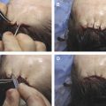
Keller and colleagues evaluated the histology, patient satisfaction, and effect on scars over 6 months post treatment with a 1064-nm laser. The 12 patients in this study had skin types II to V with mild to moderate atrophic facial scars. There was a significant increase histologically in dermal collagen after treatment. Based on pre- and posttreatment photographs, clinically there was at least moderate improvement in at least 50% of the patients. One patient with dark skin experienced PIH; however, it resolved within 4 months. The patient in Fig. 1 , who was not part of this study, has facial acne scars that were treated with a 1064-nm laser.