Because the Latino/Hispanic ethnic group is made up of various skin phototypes there is no one particular laser parameter applied to all Latinos. This review examines specific laser therapies and tailors them for usage in the Latino population. Particular emphasis is placed on the selection of laser parameters, wavelengths, and pulse durations that are suitable and safe to use in Latino subtypes. Limitations are noted in the availability of certain lasers and the cost of such treatments as well as how the phototype of the patient limits what parameters can be used.
Defining skin of color and the Latino population
Defining skin of color can be a challenge; a universal definition is not known. The terminology skin of color encompasses not only cultural aspects, but historical ones as well. There are also religious and geographic considerations to consider. The term skin of color can refer to individuals of certain racial and ethnic groups who share similar defining characteristics. Certain skin type populations are not only defined by the hues of their skin but also by the cutaneous diseases and reaction patterns that they share. Races are groups of people defined by the continent from which they originated. Although it is a matter of perspective for who is defining skin of color it is generally believed that skin of color identifies racial groups with darker skin hues other than that of white skin. This definition includes groups such as Africans, Asians, Native Americans, and Pacific Islanders. The United States is unique with its larger heterogeneous population. The US Census Bureau recognizes 5 racial categories: American Indian or Alaska Native; Asian; Black; Native Hawaiian or Pacific Islander; and White. There is a genetic basis as well for skin of color. Lamason and colleagues identified the SLC24A5 gene, which is localized to the melanosome, as the determinant of the skin pigmentation gradient between blacks and whites. West Africans possess the normal form of the SLC24A5 gene, which produces brown skin, whereas whites of European descent possess a modified SLC24A5 gene. This gene produces fewer and smaller melanosomes, which results in a white skin type.
Individuals who possess skin of color can also be defined by ethnicity. An ethnic group, or ethnicity, is a group of people whose members identify with each other through a common heritage, common language, and culture, which often includes religion and a tradition of common ancestry. Members of an ethnic group are conscious of belonging to the group and recognize the group’s distinctiveness from other ethnicities. In the United States the fastest increasing ethnic group is the Hispanic or Latino population. The term Latino was officially adopted by the United States government in 1997 to define the ethnicity as Hispanic for anyone who is of Spanish descent, speech, or ancestry. According to US Census population projections for 2050 it is estimated that the nonwhite skin of color population will equal that of the non-Hispanic white population and 29% of those will be Latinos. The US Census encompasses the Mexican, Cuban, Puerto Rican, Central American, South American, and other populations of Spanish descent as being part of the overarching Hispanic/Latino ethnic group. This ethnic group is composed of diverse populations and their skin hues can range from white to black. Correspondingly the Hispanic/Latino population is composed of various ancestries, which include white, Native Indian, African, and European descents. As a result of the eclectic makeup of this ethnic group it can be challenging to put forth strict guidelines regarding diagnosis and treatment of skin conditions that occur. Skin of color has distinct reaction patterns to cutaneous disease and this must be taken into consideration when approaching treatment options. With the rapid increase in patients with skin of color in the patient population it is imperative that the physician takes into account the cultural, genetic, and physiologic aspects of each population.
There is a paucity of research and epidemiologic information regarding the Latino/Hispanic ethnic group. Sanchez identified the top common skin disorders affecting this group. In the private and hospital-based dermatology setting acne vulgaris, eczema/contact dermatitis, photoaging, facial melasma, and hyperpigmentation ranked among the more commonly encountered problems. In contrast, a comparative practice survey carried out at the St Luke’s Roosevelt Hospital Skin of Color Center in New York City, between 2003 and 2004, revealed that the top most common skin disorders in the black population are: acne vulgaris, dyschromia, contact dermatitis, and alopecia and seborrhea dermatitis. These contrasting epidemiologic results show that within the realm of skin of color, all are not the same and a “1 treatment fits all” policy cannot be applied.
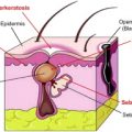
Although there is a paucity of medical knowledge on the differences between skin structure and function between racial and ethnically distinct skin, differences do exist. Reed and colleagues showed that skin phototypes V and VI, when compared with skin phototypes II and III, required more tape strippings to disrupt the epidermal barrier. This finding implies that the more compact cornified skin layers in the darker skin types displayed increased epidermal barrier function and a quicker recovery time. This finding was not true of white skin compared with Asian skin, showing that this inherent property was related to skin phototype and not race. In a study by Sugino and colleagues the ceramide content variability of different skin phototypes was measured. Ceramides are the major lipid constituent of lamellar sheets present in the intercellular spaces of the stratum corneum. These lamellar sheets are believed to provide the barrier property of the epidermis. Therefore externally supplied ceramides function by incorporating into the intercellular lipid of stratum corneum to replace the depletion that occurs with aging and environmental damage such as from surfactant exposure. Topical ceramide has been shown to improve barrier function of damaged skin both acutely and chronically, as assessed by reduced transepidermal water-loss assays. This improved barrier can reduce skin sensitivity and responsiveness to environmental insult, leading to reduction in skin appearance problems such as redness and potentially the manifestations of aging. The lowest ceramide levels were in black skin, followed by whites, Hispanics, and Asians. Ceramide levels are directly proportional to water content and inversely proportional to transepidermal water loss. Whether these structural differences lead to functional differences for patients of Latino/Hispanic skin type is still under investigation, with more rigorous studies needed to assess irritability and erythema induction in skin of color.
Melanocytes within the epidermis are an important structure when assessing differences in skin of color. Melanocytes are derived from neural crest cells and migrate into the basal layer of the epidermis. These cells transfer melanosomes containing melanin into neighboring keratinocytes. One of the main functions of melanin is to provide ultraviolet (UV) radiation protection. It is known that melanocyte number is constant among all races. The differences in clinically perceived skin color depend on the amount, density, and distribution of melanin within the melanosome and inherent melanocyte activity. Therefore the differences in melanosome density, size, and aggregation correlate with the differences in skin pigmentation. Fair-skinned individuals have more stage I and II melanosomes, which are small, more aggregated, and degraded quicker in the stratum corneum compared with darker-skinned individuals who have stage IV melanosomes, which are larger, nonaggregated, and degraded at a slower rate. In skin of color, the higher number of larger, nonaggregated melanosomes correlates with a higher absorption of UV light. Darkly pigmented skin has average minimal erythema doses 15 to 33 times greater than that of white skin, with variation depending on skin tone. The melanin pigment in black skin is a neutral density filter, which reduces all wavelengths of light equally. This is important to consider when treating skin of color, especially Latino skin, which can run the gamut of phototypes.
The dermis is a highly vascular structure, which is compromised of multiple components and houses appocrine and eccrine glands. Darker skin types compared with lighter phenotypes have a thicker and more compact dermis with smaller collagen fiber bundles, more numerous, larger, and multinucleated fibroblasts, and numerous and larger melanophages. There is greater eccrine gland activity when comparing black patients with white patients, with Hispanic patients possessing gland activity between that of black and white eccrine gland activity. All these structural differences lead to important implications in assessing and treating skin of color, especially the Latino population. In the epidermis the increased melanin content implies a lower rate of skin cancer overall; however, the risk and rate of skin cancer is still real. The increased melanosome dispersion implies less pronounced photoaging. However, also because of this situation the pigmentary disorders that do arise in skin of color are a result of both biologic inheritances and cultural practices. The higher rate of pigment alteration after adulteration of skin of color is also concerning and therefore must be addressed beforehand. In the dermis the more multinucleated and larger fibroblasts correlate with the greater incidence of keloids experienced in darker phototypes.
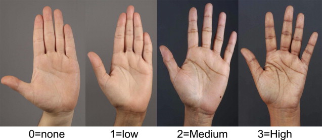
With the Hispanic population being the fastest increasing population in the United States, treating people of this ethnic/racial grouping is a growing priority. Although the census may group certain ethnic backgrounds into an encompassing category of Hispanic, biology does not. Therefore when treating a patient who identifies themselves as Hispanic, the keen physician must accurately assess and tease out the phototype and phenotype of that patient’s skin. The Hispanic population encompasses the range of phototypes and therefore 1 rule cannot apply to all Latinos/Hispanics. Dr Leal, dermatologist in an academic center in Monterrey, Mexico, has tried to decipher the best way to predict the propensity of white phototype Latinos to postinflammatory hyperpigmentation. His studies have revealed that pigmentation of palmar and digital creases is the best predictable factor for postinflammatory hyperpigmentation in Latinos. Dr Leal and colleagues (personal communication pending manuscript publication, 2010) has divided the color diversity of the palmar and digital creases into 0 to 3, with the higher number indicating a darker skin tissue response despite skin phototype ( Fig. 1 ). With a solid understanding of laser physics and properties and tissue interactions dermatologic lasers can be used in all patients regardless of skin color. Safe use of these lasers strongly depends on proper selections of laser wavelengths and suitable laser parameters. Some overarching principles can be applied to all patients undergoing a dermatologic laser procedure. Test spots are strongly encouraged in all patients with darker and even lighter phenotypes with dark palmar and digital creases, allowing the physician and patient to assess the efficacy and safety of the specific laser before undergoing a complete procedure. This strategy also inadvertently strengthens the physician-patient bond and establishes a trusting relationship. Also, preoperative expectations and goals need to be communicated and the inherent risks and complications need to be addressed beforehand. When treating patients with darker phenotypes less aggressive parameters are recommended to minimize unwanted adverse effects, with the realization that multiple treatments are usually necessary in patients with darker phototypes.