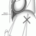
Fig. 49.1
To control injuries at the base of the mesentery, begin by placing the root of the mesentery between the index and middle fingers of the nondominant hand
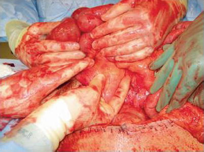
Fig. 49.2
Next, place the thumb and index finger on opposite sides of the injury. By squeezing the area of injury in this fashion, effective hemostasis is quickly achieved. The sheets of mesentery can then be incised and the points of bleeding accurately identified and addressed
If the injury is to the bowel itself, at this point in the operation you should be looking at several rapidly closed holes with long silk tags, or areas of hematoma adjacent to the colon or at the bowel/mesentery junction without frank spill. The first step is to avoid the temptation to start contending with the holes that you know you have and instead fully define all of the injuries with which you are dealing. This may save you from wasting time on primary repairs that will later wind up in a resected piece of bowel after other injuries are discovered. While this is frequently straightforward, a few particulars are worth mentioning. First, any pericolonic hematoma needs to be completely inspected. This means rolling the colon and dissecting all other tissues away until only the colonic serosa remains. If a subserosal hematoma is found, unroof it. Similarly, any hematoma at the junction of the bowel wall and mesentery should be considered to be an injury until proven otherwise by direct visual inspection of the serosa of the involved area. Do not let the fear of an iatrogenic injury stop you from completely inspecting the area of concern. Occasionally, for injuries close to the root of the mesentery, it will be necessary to actually take the ligament down. The important thing to remember is to never look at a hematoma immediately adjacent to a viscus and leave it thinking that it is probably OK. In the words of the esteemed surgeon Benton DuPont, “That works about as well as ball bearing book ends.” Similarly, an odd number of holes in the bowel should trigger fears for a missed injury. While tangential wounds can occur, the finding of an odd number of holes should cause one to spend extra time looking for a missed perforation. Keep in mind the mobility of the small bowel, that people get shot in all manner of bodily positions, and that bullets do not travel in straight lines. In short, when looking for bowel injuries, paranoia is a healthy attitude.
Once an injury to the bowel wall is located, debriding the edges to healthy tissue and a transversely oriented primary repair should be the default first choice. Personally, we prefer a single-layer running suture. If we have a relatively short segment of the small bowel that has been fluted, we will resect it in order to minimize the number of suture lines. When making the decision about whether or not to resect an intervening piece of small bowel between two injured segments in order to save an anastomosis, we will check to see if we are leaving the patient with at least 250 cm of small bowel if a resection is performed as short bowel syndrome should not be a concern with that length. In adults, particularly those lacking a functional colon, lifelong TPN dependence is likely to occur in those who have 100 cm or less. While the presence of a functional ileocecal valve is thought to increase this length to an unknown degree, trauma patients are clearly at higher risk for subsequent bowel resections at their initial admission as well as over their lifetime (trauma recidivism rates are significant, proving the old saying, “Trauma is a chronic disease with acute exacerbations”). Given that, it is smart to leave them with plenty of bowel to spare. When dealing with two enterotomies that are within a centimeter or so of each other, the bridging wall of bowel separating them can become devascularized and slowly necrose over the course of a few days. Avoid the temptation to perform two primary repairs in this setting, as it is prudent to debride this segment and convert them into one larger enterotomy which can then be closed easily.
If a resection is required, the decision to perform an anastomosis versus diversion has vexed generations of surgeons. During World War II, failure to treat a penetrating colon injury with diversion was a court martial offense. With time, as it came to be realized that civilian injuries are not necessarily equivalent to military injuries and that ostomy reversal carried its own morbidity, the pendulum began to swing toward a lower threshold for anastomosis at the time of the first operation. In the stable, previously healthy penetrating trauma patient who is exhibiting no signs of shock and has received no blood products, we will always perform a primary anastomosis regardless of the location of the injury or the amount of peritoneal soilage from stool. This includes left colon injuries. The one exception to this general rule is the elderly patient with minimal physiologic reserve. In that setting, we will still perform enteroenterostomy and enterocolostomy. When considering the higher risks of colocolostomy in conjunction with the fact that an anastomotic leak in this specific population is often a death sentence, we will generally opt to perform resection and diversion in those patients. One should realize though that frequently means these will become permanent ostomies.
Typically the stable patient has relatively normal caliber bowel, and if this is the case, either hand-sewing or stapling the anastomosis should be fine.
49.3 The Second Pass-Through the Abdomen: Damage Control
Now let us consider a different situation. You have just finished a fast right nephrectomy and packing the liver; a quick glance up shows you multiple units of blood products hanging on the IV pole and on the floor around anesthesia’s side of the drapes. At this point, anesthesia tells you that the patient’s core temperature is 33.9 °C. You need to be getting out of this patient’s abdomen ASAP. Having temporized the patient’s enterotomies on the first pass with whip stitches, what do you do now?
For the patient who is in extremis, the answer is simple: nothing. Leaving the injuries alone with figure-of-eight closures is not something about which you should be cavalier, because this suboptimal technical closure is prone to breaking down (particularly in the setting of bowel edema and splanchnic vasoconstriction from shock and pressors). In the patient who is actively dying, however, the risk posed by this strategy may be balanced out by the benefit of saving a few minutes in a setting where time is critical. If one elects to leave the whip stitches in place, it is important that you take the patient back at or around 24 h post-injury as longer delays begin to make the risks of breakdown with renewed spill of stool prohibitive. While we realize that the plural of anecdote is not data, the senior author has resorted to this strategy many times and has yet to have a whip-stitch closure break down in this time frame.
If a damage control approach is being utilized but the patient is not in extremis, a different approach is used for hollow viscus injuries temporized on the first pass-through the abdomen. If it is a matter of dealing with three or fewer small, discrete perforations, we will take the time necessary to perform simple, running, single-layer closures. These definitive repairs are quick, and a time-consuming second layer is unnecessary. If many more injuries exist, or a destructive injury is present, one should resect the involved piece of bowel and leave the patient in discontinuity. Alternatively, one can fire a linear cutting stapler adjacent to both sides of an injury, effectively excluding the injury from the fecal stream and preventing spill. With a functioning nasogastric tube, patients tolerate being left in discontinuity surprisingly well and can be left in this fashion for 48 h or longer if circumstances require it. Diversion in this setting is unnecessary and in fact can be problematic as the abdominal wall frequently becomes quite edematous in damage control patients. This can put an ostomy that initially looked good under significant tension and predispose it to ischemia.
Related posts:

Stay updated, free articles. Join our Telegram channel

Full access? Get Clinical Tree


