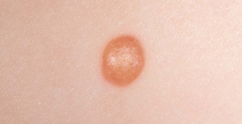
Juvenile xanthogranuloma


Juvenile xanthogranuloma


Megan Mowbray and Olivia M.V. Schofield


Evidence Levels:  A Double-blind study  B Clinical trial ≥ 20 subjects  C Clinical trial < 20 subjects  D Series ≥ 5 subjects  E Anecdotal case reports


image
Courtesy of Mary Glover, Great Ormond Street Hospital, London, UK


Juvenile xanthogranuloma (JXG) is a benign disorder characterized by solitary or multiple, yellow-red nodules in the skin, and, occasionally, in other organs. The lesions spontaneously regress by age 6 months to 3 years. It occurs most commonly in infancy and early childhood, but adults may be affected. Treatment is only recommended for systemic lesions if their location interferes with organ function.


Management strategy


The diagnosis of JXG is usually made clinically, but a biopsy is required for atypical clinical variants (giant, plaque-like, paired, clustered, infiltrative, lichenoid, linear, subcutaneous, and intramuscular) and if multiple lesions are present. JXG is classified as a ‘non-Langerhans cell histiocytosis’ and characterized histologically by an accumulation of foamy histiocytes lacking Birbeck granules (non-Langerhans cells) and multinucleated Touton-type giant cells. Immunohistochemical staining of JXG lesions shows expression of factor XIIIa, CD68, CD163, fascin, HLA-DR, and CD14, but not S100 or CD1a. This pattern of expression suggests that the cell of origin of JXG is the dermal dendrocyte. The histogenesis of this group of conditions is unclear but the relationship to LCH is thought to be a divergent differentiation from a common precursor CD34+ myeloid cell. Reported cases of the two conditions occurring in the same individual suggest that this divergent maturation is not irreversible.


The ‘JXG family’ can be divided into three groups:


In the most common solitary type of JXG (60–89% of all cases), there is a male to female preponderance of 1.5 : 1. The usually affected sites are the head, neck, and upper trunk. In young infants with multiple lesions the male : female ratio increases to 12 : 1. In the majority of cases no further investigation is required. The lesions will resolve spontaneously within months or years and no follow-up is necessary. As involvement of other organs can occasionally occur in the cutaneous group, a review of systems and general examination is recommended to look for involvement of other sites and to examine for café-au-lait macules (see below).


Specific investigations


Stay updated, free articles. Join our Telegram channel

Aug 7, 2016 | Posted by in Dermatology | Comments Off on Juvenile xanthogranuloma

Full access? Get Clinical Tree

Get Clinical Tree app for offline access