Introduction to Vulvar Disease
VULVAR ANATOMY (Figures 1.1., 1.2., 1.3., 1.4., 1.5., 1.6. and 1.7., Vulvovaginal Consultation Form)
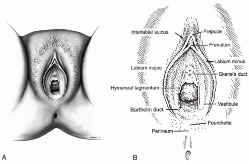
An understanding of the normal external anatomy of the vulva is of great value in recognizing pathologic changes, as well guiding the pathologist to the appropriate differential diagnosis should a biopsy be obtained. The epithelium of the vulva is highly variable, as are the types of subcutaneous tissues and accompanying skin appendages, if present. The key anatomic sites of the vulva are illustrated in Figure 1.1.A.
DEFINITION
The vulva is composed of the vestibule, the clitoris, the labia minora, the labia majora, and the mons pubis. The vestibule is that portion of the vulva that extends from the exterior surface of the hymen to the frenulum of the clitoris anteriorly, the fourchette posteriorly, and laterally to the Hart line, where nonkeratinized squamous epithelium of the vestibule meets the keratinized epithelium of the more lateral labia minora (Fig. 1.1.A.). This stratified nonkeratinized squamous epithelium has a thickness of approximately 1 mm. Within the vestibule are found the urethral orifice, openings of the Skene ducts, openings of the Bartholin ducts, openings of the minor vestibular glands, and the vaginal introitus.
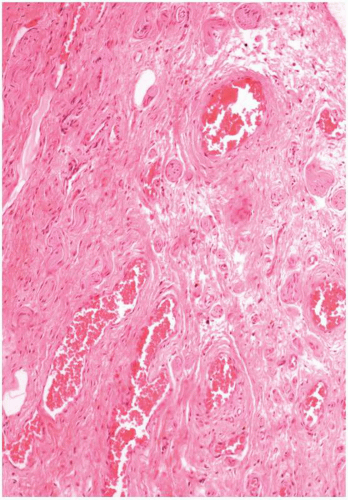
 Figure 1.3. Vulvar vestibule, postmenopausal woman. The epithelium is thin and nonkeratinized and has mild spongiosis. The subepithelial tissue contains many small vessels and is collagen rich. |
THE MONS PUBIS
The mons is that portion of the vulva presenting as the rounded fleshy prominence over the symphysis pubis. The mons epithelium is composed of stratified squamous epithelium that is hair bearing. The skin of the mons is similar to that of the labia majora, with pilosebaceous units distributed throughout its substance. Hair follicle depth may be up to 2.72 mm. The underlying tissue of the mons is composed primarily of adipose tissue.
THE VULVAR VESTIBULE
The vulvar vestibule is the portion of the vulva extending from the exterior surface of the hymen to the
frenulum of the clitoris anteriorly, fourchette posteriorly, and Hart line laterally. The vestibule is thus bounded by the junction of the nonkeratinized squamous epithelium of the vestibule junctions with the keratinized epithelium of these peripheral sites, such as the more papillated appearing keratinized epithelium of the more lateral labia minora (Fig. 1.1.B.).
frenulum of the clitoris anteriorly, fourchette posteriorly, and Hart line laterally. The vestibule is thus bounded by the junction of the nonkeratinized squamous epithelium of the vestibule junctions with the keratinized epithelium of these peripheral sites, such as the more papillated appearing keratinized epithelium of the more lateral labia minora (Fig. 1.1.B.).
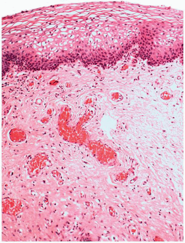
Stratified nonkeratinized squamous epithelium forms a mucosa with a thickness of approximately 1 mm (Fig. 1.2.). The nonkeratinized squamous mucosa of the vestibule is glycogenated in women of reproductive age, or under estrogen influence, and resembles vaginal mucosa.
Structures within the vulvar vestibule include the urethral orifice, openings of the Skene ducts, vaginal introitus, opening of Bartholin ducts, and linea vestibularis. The linea vestibularis is observed in approximately one quarter of newborn female infants and is located in the posterior portion of the vestibule. It is a white streak or spot in the midline of the posterior vestibule that extends nearly to the posterior commissure.
THE CLITORIS
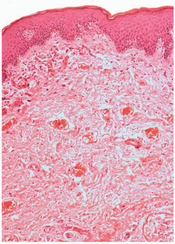
The clitoris is homologous to the corpus cavernosum of the male penis. The clitoris consists of two crura and a glans clitoris. The crura are composed of erectile tissue that is enveloped by the tunica albuginea (Fig. 1.4.). The clitoris is attached bilaterally to the labium minus by the frenulum and has a length of 16 ± 1.4 mm, with a transverse diameter of 3.4 ± 1.0 mm and a longitudinal diameter of 5.1 ± 1.4 mm in adult women (Fig. 1.1.B.). Although height and weight do not influence size, parous women tend to have larger measurements (Verkauf et al, 1992).
The epithelium is squamous mucosa without glands, rete, or dermal papillae. The clitoris is rich in sensory receptors, as are the labia minora and majora.


