94 Instrumentation and Supplies Used in Hair Restoration Surgery
Summary
Keywords: instrumentation supplies hair restoration surgery donor harvesting site creation blades forceps needles microscopes blades
Key Points
•There is a great variety of instruments. Choose what you think will be best for you and focus on technique.
•Begin wisely and talk to experienced colleagues in the field of hair restoration surgery.
•Listen to your surgical assistants. They can also help you to choose the right instruments.
•New instruments are developed every year. Some can really improve your daily routine. Keep updated.
94.1 Introduction
One of the first steps a physician wanting to perform hair transplantation surgery must take is obtain the proper instruments and supplies. Instrumentation continues to improve every year, making the surgeon’s work easier and the results better. Both common and specialized instruments are used in hair transplantation. Many choices exist with subtle variations, each claiming to be better than the other.
It would be impossible to list and describe all the different instruments and supplies used in hair transplantation. However, we can give an overview of the essential instruments and supplies required in the multiple stages of modern hair restoration surgery as well as discuss various alternative instruments for the different techniques.
Many of the surgeon and staff choices of instrumentation will be based on personal preference after a trial of different devices. The following information is a compilation of both the authors’ combined experience of greater than 30 years as well as discussions with peers.
94.2 Analysis of Recipient and Donor Areas
Evaluating the donor area and recipient area is a critical first step for making a surgical plan. The donor area needs to be evaluated to determine its size, laxity, donor density (FU/cm2), and hair characteristics such as caliber and percent miniaturization. The goal is to be able predict the amount and quality of donor hair that can be safely harvested. The recipient area needs to be evaluated for size and severity of the current areas of balding and percent miniaturization in potential balding areas. The goal is to determine the number of grafts needed to treat certain areas and predict the risk of future hair loss.
Donor laxity can be estimated simply using one hand, but a tool such as the Scalp Laxometer (www.georgetiemann.com) can also be used.
Donor density, hair caliber, and percent miniaturization can be evaluated by various imaging devices with different levels of sophistication.
•The least sophisticated are simple densitometers like the Welch AllynTrichoscope or Eschenbach densitometer.
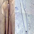
•More advanced digital or analog dermatoscopes exist such as the Micro-Vid (www.micro-vid.com), Folliscope (www.hairscience.co.kr), Dino-lite (www.dino-lite.com) or Dermalite (www.dermalite.com; Fig. 94.1). These devices enable the creation of high-resolution macrophotographs for evaluation. Some come with their own software for scalp and hair evaluation, while others use third-party software such as Canfield Imaging software (www.canfiieldscientific.com).
•Complete sophisticated imaging systems with software analysis exist as Fotofinder (www.fotofinder.com; Fig. 94.2). These may be overkill for a general practice but are ideal for research.
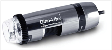
Fig. 94.1 Videomicroscopy.
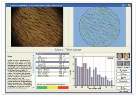
Fig. 94.2 Fotofinder Trichoscale Trichoscan.
The sizes of the donor and recipient areas are usually measured with a simple tape measure or grids and templates that can overlay the scalp.
94.3 Preoperative Patient Preparation
The items listed in Table 94.1 are useful in preparing the patient before surgery. Various marking pens or pencils are used to plan the recipient area, especially the hairline. Scissors or hair clippers are used to trim the planned donor area. A comb, tape, lint brush, or handheld vacuum can be used to remove loose hairs. It is useful to have a spray bottle with normal saline or aqueous disinfectant to wet and clean the donor and recipient areas prior to surgery.
Table 94.1 Preoperative preparation setup
●Marking pen or wax pencil |
●Scissors or hair clipper |
●Hair comb, lint roller, or portable vacuum to remove hair |
●Surgical ruler and or compass to take measurements |
●Handheld mirror |
●Tape, hair clips, and hair bands to keep hair out of the way |
As many procedures are carried out under both sedation and local anesthesia, preoperative assessment, and intraoperative monitoring of pulse, oxygen saturation, and blood pressure should be regarded as mandatory. Various oximetry (Fig. 94.3) devices such as the Welch Allyn monitor are available. In addition, an automatic external defibrillator (AED; Fig. 94.4) should be available for emergency use as well as oxygen and resuscitation equipment (see Chapter 33).
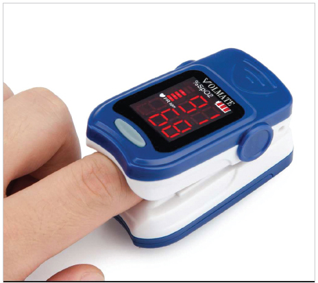
Fig. 94.3 Digital oximeter.
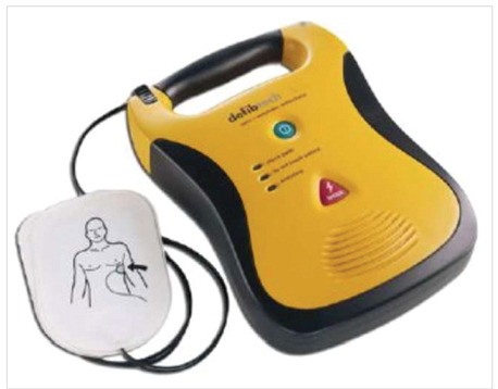
Fig. 94.4 Automatic external defibrillator (AED).
94.4 Sedation, Anesthesia, and Tumescence
Many surgeons use oral or parenteral sedation, commonly with benzodiazepine variants such as midazolam or diazepam. In addition, some surgeons use narcotic analogs. Differing regulations in various jurisdictions regarding the requirements for intravenous sedation, if used, should be followed.
Local anesthesia is most commonly performed using lidocaine, sometimes together with bupivacaine, in combination with adrenaline. Apart from normal syringe and needle devices, advanced injection instruments such as the CompuMed Wand (for controlled injection of anesthetic solution) or the Uni-Matic syringe or (for controlled tumescent injection) may be useful. The Self-Filling Injector device (Cole Instruments) is another instrument often used for injecting tumescent solution.
Ancillary devices such as a handheld vibrator are also commonly used to reduce the discomfort of injection of local anesthetic (Fig. 94.5; see Chapters 31 and 58).
Fig. 94.5 Handheld vibrator.
94.5 Donor Harvesting
Modern donor harvesting involves two differing techniques: donor strip harvesting (follicular unit transplantation [FUT]) and follicular unit excision (FUE). The dominant technique is still strip harvesting although FUE is rapidly gaining in popularity. The instrumentation is different depending on the approach you take.
94.5.1 Follicular Unit Transplantation/Strip Surgery
Our basic instrument set used for strip harvesting is shown in Fig. 94.6. Surgeons typically perform strip harvesting with a single scalpel blade using a no. 10blade or a no. 15blade.
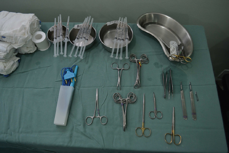
Fig. 94.6 Surgical setup.
Related posts:
 Hair Anatomy and Histology for the Hair Transplant Surgeon
 Plugged in: How to Ensure That Your Practice Thrives (and Survives) in Today’s DigitalWorld
 Transplanting into Areas of Cicatricial Alopecia
 Special Considerations for Postoperative Care in Follicular Unit Excision
 Hairline and Recipient Area Repair of Poor Previous Transplantation
 Ergonomics in Hair Restoration Surgery: FUE Technique
Stay updated, free articles. Join our Telegram channel

Full access? Get Clinical Tree


