SKIN BARRIER INTEGRITY:
From Algal Protective Exoskeleton to a Protective
Barrier for the Epidermis
Author
Alexandra Jeanneau
Scientific communication officer,
Alban Muller Group
ABSTRACT
The skin epidermis serves numerous functions, but its primary purpose is to act as a protective barrier for the body from the external environment. It is composed of keratinocytes piled in layers lying upon each other thanks to specific complex junctions maintaining cohesion and communication among them. With aging this “young” architecture becomes disorganized, and this leads to a weaker protection and poorer communication of information signals between cells.
Some living organisms have developed a protective mechanism to this disorganization by an adaptation to their environment. For instance, Padina pavonica, a brown alga, builds an organized external deposit of calcium carbonate in the form of aragonite crystals for its protection.
As calcium bioavailability is necessary for the synthesis of essential epidermis structures, a high-tech active extract of Padina pavonica was developed and studied. This study highlights the activity of the extract for maintaining bioavailable calcium and thereby stimulating the synthesis of calcium-depending structures, crucial for the epidermis-protective function.
4.1.6.1 Skin barrier and epidermis organization overview
a. Epidermis cell structures for skin resistance and cohesion
b. Cell structures depending on calcium
c. Calcium’s information-signaling properties
4.1.6.2 Solution for alterations in epidermis
a. Epidermis structures responsible for epidermis resistance
b. Improving calcium bioavailability
a. A primitive strategy involving calcium
b. An ingredient with a unique mode of action
c. Stimulation of calcium-depending cell structures
d. Restoring intercellular communication, a key factor of
the epidermis functioning
e. Improving skin barrier integrity: a better protective
shield against pollution
INTRODUCTION
The primary function of the epidermis is to act as a protective barrier for our body from the external environment. Maintaining its integrity and resistance is crucial for proper functioning of the skin. In view of this function, and our present understanding of it, there is considerable ongoing research on skin to improve its strength and to find new active ingredients with innovative modes of action that improve its integrity.
4.1.6.1 SKIN BARRIER AND EPIDERMIS
ORGANIZATION OVERVIEW
Skin barrier function resides primarily within the stratum corneum (SC), the top layer of the epidermis. It plays a vital role in forming a protective barrier and helps to prevent percutaneous entry of harmful pathogens in the body.
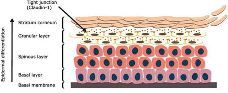
Epidermis is the upper layer of the skin tissue, just below the stratum corneum. It is composed of several layers of keratinocytes that undergo continuous morphological changes reflecting their underlying keratinization as a protective barrier of the epidermis (both mechanical and chemical).
This epidermal differentiation begins from the depth to the surface of the epidermis cells and enables the distinguishing of four layers on a skin cut: the germinal layer (or basal), the spiny layer (or spinous), the granular layer and horny layer, or stratum corneum (first compacted then desquamating). Each stage of epidermal differentiation is characterized by the expression of specific proteins.
Therefore, maintaining a good level of proliferation and differentiation of keratinocytes from within the epidermis is crucial for the proper functioning of the skin barrier and for its integrity. Thus, the most important function of keratinocyte differentiation is the establishment and maintenance of the stratum corneum, since the latter consists of corneocytes and extracellular lipids secreted by differentiated keratinocytes, which together function as a barrier against the entry of chemicals and microbes from the environment and protect the body from dehydration (Figure 1).

a. Epidermal cells structures for skin resistance and cohesion
The mechanical strength of the epidermis depends on the integrity of the epidermal cell skeleton, which primarily consists of cytokeratins. Keratinocytes are piled in layers of cells, lying on each other, in which cytokeratins are synthesized in high quantities. Cytokeratins are specific proteins building the cytoskeleton of keratinocytes and responsible for their resistance. In each layer, daughter cells only differ from mother cells by the maturation of cytokeratins, which undergo modifications from the deepest layer of the epidermis to its surface. The most external layers are made of corneocytes (final form of the keratinocytes’ differentiation) that no longer contain a nucleus and are filled with almost only a framework of modified cytokeratins.
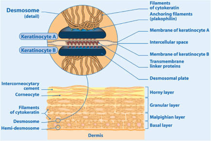
The structural integrity of the skin also depends on dynamic junctions between cells called desmosomes. They are special membrane-associated structures of the epidermis equivalent to press studs that attach or detach depending on the cell needs. Indeed, the formation of desmosomes mostly takes place in the basal layer, the deepest one in the epidermis, where it is favored by the palissadic arrangement in perpendicular layers of keratinocytes placed very near one to the other. Having a particular structure at the junction of the basal layer with the dermis, where they are called hemi-desmosomes, desmosomes evolve from one layer to the other to end up in corneo-desmosomes at the stratum corneum level, where enzymes break them to favour the desquamation process.
Desmosomes are highly organized and build an intercellular adhesion point strongly anchored by cytokeratin filaments that penetrate deeply into the cytoskeleton, thus ensuring cohesion for the functioning of the epidermis.
The resulting “clipped” architecture of the epidermis explains its resistance and cohesion but also favors good intercellular signals, which are necessary for its proper function (Figure 2).

b. Cell structures depending on calcium
Indeed, desmosomes and cytokeratins are closely associated with each other, ensuring the junctions between keratinocytes and the cohesion between epidermis and the underlying dermis. It is therefore important to emphasize the critical role of both structures for the consolidation of the mechanical function and the integrity of the epidermis.
Cell communication is ensured by chemical messengers passed from one cell to another, a process that also depends on desmosomes and cytokeratins cell structures, which are calcium-dependent.
Indeed, calcium is involved in the maturation of cytokeratins as well as in the adhesion of the desmosomial proteins. Calcium ions are transferred from the blood to the most superficial cells by channels specialized in ionic transport in cell membranes. Calcium is then attracted to the structures on which it must act, particularly cytokeratins and desmosomial proteins.
c. Calcium’s information-signaling properties
Calcium is an excellent information aid, as it can combine with a number of acids and be included in many minerals. The same salt with the same chemical formula can result in different minerals, depending on the atomic arrangement. Indeed, oyster shell, for instance, is covered by an outer layer, the thickest one, made out of an amorphous (thus nonorganized kind of calcium carbonate) and a thinner layer of shining calcite, itself covered with mother-of-pearl; the two latter minerals are crystallized (thus organized) forms of calcium carbonate. The soluble form of these minerals releases calcium ions with distinct solubility coefficients in the surrounding media and are able to pass on electric potentials at different levels. This process allows for the circulation of information. In mammals, the role of calcium is essential for blood coagulation, muscle contraction, nervous influx transmission, and heart rhythm regulation.
Signaling information associated with calcium in the skin is observed at two levels: the maturation of cytokeratin proteins that set up mechanisms of apoptosis, program cells’ death for protection purposes (renewal of the epidermis), and the organization of skin’s immune defense. Indeed, the terminal differentiation of epidermal keratinocytes, linked to the maturation of cytokeratins, is an essential process for the renewal of keratinocytes and ends in the formation of enucleated “dead” cells: the corneocytes. Therefore, calcium is a key regulator in epidermis homeostasis of keratinocyte proliferation and differentiation.
Many studies highlight the signaling function of calcium ions on keratinocytes as promoting cell-cell adhesion, and differentiation-related genes. Some studies (1–2) provide evidence that keratinocytes deficient in the calcium-sensing receptor fail to respond to calcium stimulation and to differentiate, indicating a role for the calcium-sensing receptor in transducing the Ca2+ signal during differentiation. It has also been observed that calcium is involved in recovery and wound-healing. Immediately after a scratch, there is a wave of increased calcium ionic transport outward from the damaged area that remains elevated in a peripheral layer of cells around the damaged area for several minutes and then disappears gradually. These studies confirm a signaling pathway that appropriately directs the wound-healing process in epidermis (3). Calcium signaling via gap junctions might be involved in the induction of calcium waves in response to mechanical stress at the upper layer of the epidermis (4).
4.1.6.2 SOLUTION FOR ALTERATIONS IN EPIDERMIS
Stay updated, free articles. Join our Telegram channel

Full access? Get Clinical Tree


