Inferiorly Based Skin-Muscle-Conjunctival Advancement (Cutler-Beard) Flap From The Lower to The Upper Eyelid
C. BEARD
J. H. SULLIVAN
EDITORIAL COMMENT
In using so much of the full-thickness skin and conjunctiva of the lower lid to recreate the upper lid, the danger of a significant portion of patients developing ectropion is something to be considered before selecting this procedure for use.
The upper eyelid is far more important functionally than the lower lid, since it is responsible for almost all the protection of the cornea. Without an adequate upper lid, the globe cannot survive long as a useful organ. Reconstructive procedures for major upper eyelid repair are few and complex. The inferiorly based skin-muscle-conjunctival advancement flap has withstood the test of time. This two-stage procedure can be used for total lid replacement, yet it is one of the simplest of the upper eyelid reconstructive operations.
INDICATIONS
The advantages of this procedure are its usefulness in correcting large upper lid defects, its simplicity, and the surprising freedom from deformation of the lower lid (1). The necessity for prolonged visual obstruction with this procedure may be a problem in an occasional patient. A much shorter period of occlusion is possible with the Mustardé “switch flap” (2). It may be possible to reduce the period of occlusion thought to be necessary with the Cutler-Beard flap, but we have not attempted to do so.
ANATOMY
This flap consists of an inferiorly based full-thickness lower lid flap (skin, orbicularis muscle, orbital septum, lower lid retractors, and conjunctiva) advanced beneath the lower lid margin to replace the upper lid.
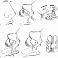
OPERATIVE TECHNIQUE
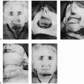
The procedure (Fig. 24.1) begins with complete removal of the upper lid tumor under frozen-section control or freshening of the margins of traumatic or congenital defects. Remnants of the remaining lid are held under moderate traction to lessen the width of the defect. The measurement of this width is used to determine the width of the full-thickness lower lid flap. A horizontal skin incision of the same distance is made. It is deepened at one end into the inferior fornix. Using scissors, a full-thickness horizontal incision is completed immediately below the tarsus to the width of the skin incision. Full-thickness vertical scissor cuts are made from the ends of the horizontal cut to the extreme depths of the inferior fornix, forming a rectangular flap. The flap is mobilized by undermining the skin inferiorly over the orbital rim. It is then advanced posterior to the bridge of the lower lid margin into the upper eyelid defect, where it rests without tension.
Related posts:
 Cheek Rotation Skin (Mustardé) Flap to The Lower Eyelid
 Wraparound Cartilage Flap for Correction of Cleft-Lip Nasal Deformity
 Oral Mucosal Flaps for Septal Reconstruction
 Postauricular and Retroauricular Scalping Flap (The Paras Flap)
 Scapular and Parascapular Flaps
 Platysma Musculocutaneous Flap to The Lower Lip
Stay updated, free articles. Join our Telegram channel

Full access? Get Clinical Tree


