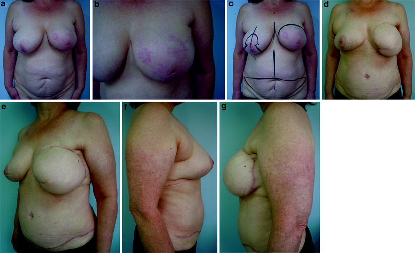
Fig. 42.1
Preoperative (a, b) and postoperative (c, d) views of a 62-year-old patient with a local recurrence after breast-conserving therapy 8 years earlier in the left breast. c, d Twelve months after one-stage breast reconstruction with an implant and contralateral breast reduction for symmetry
42.3 Flaps
The description in 1977 of the latissimus dorsi (LD) musculocutaneous flap for breast reconstruction introduced another important option for autologous tissue reconstruction in patients after mastectomy [14]. Until the description of the transverse rectus abdominis muscle (TRAM) flap in 1982 [15], use of autologous tissue for reconstructions was closely linked with breast implants. The TRAM flap provided a relatively easy technique for acquiring ample tissue for shaping and skin coverage in most reconstructions. When a large amount of skin replacement is required, it is the preferred technique.
Complications in previously irradiated patients ranged from a mastectomy defect with minimal radiation changes to frank skin necrosis. Coverage is the primary purpose for the latter group, and the reconstructive operation becomes an aesthetic procedure in the former. For the reconstructive surgeon, there are two major areas of concern after radiotherapy:
1.
The recipient bed
2.
The flap’s vascular pedicle
Breast reconstruction with a TRAM flap after radiotherapy is reasonable and should remain the first choice for most patients, although multivariable logistic regression analysis showed both obesity and prior radiotherapy to be associated with an increased risk of fat necrosis [16].
The bipedicled flap should be used when possible to allow there to be sufficient tissue for reconstruction after resection of the irradiated recipient site and provide improved blood supply to a vascular impoverished recipient bed [16]. However, using a bipedicled flap in the irradiated patient does not prevent the occurrence of fat necrosis. The rate of fat necrosis suggests some compromised blood flow to the subcutaneous fat, possibly from partial obstruction of the internal mammary artery.
The largest review of irradiated patients undergoing TRAM flap reconstructions supports previous histologic studies that large vessel damage from radiation is rare and not prohibitive for using pedicles for flaps [16]. Moreover, Kroll et al. [17], using four independent observers, compared 82 patients with a history of previous chest-wall irradiation with 202 nonirradiated patients in order to determine whether prior irradiation was associated with more frequent complications. Both groups underwent LD and TRAM flap breast reconstruction. The complication rate in the irradiated group was 39 versus 25 % in the nonirradiated group (p = 0.03). In the irradiated group, complications were more frequent with the LD flap (63 %) than with the TRAM flap (33 %; p = 0.063), but this was not statistically significant.
Although only irradiated groups were evaluated, Schuster et al. [18] in a study with patient questionnaires found higher satisfaction rates with TRAM flap reconstructions than with LD flaps or implants in previously irradiated patients (Fig. 42.2).


Fig. 42.2
Preoperative (a–c) and postoperative (d–g) views of a 59-year-old patient with an extensive local recurrence after breast-conserving therapy 4 years earlier in the left breast. d–g Twelve months after one-stage breast reconstruction with a bipedicled transverse rectus abdominis myocutaneous flap and contralateral breast reduction for symmetry
42.4 Effects of Radiation on the Decision for Immediate Breast Reconstruction
Issues concerning breast reconstruction in patients who have had or may potentially require radiotherapy include:
Effect of radiotherapy on soft tissues
Timing of irradiation in the patient presenting with breast cancer
Choice of a breast reconstruction option that will produce the optimal long-term cosmetic outcome.
The effects of radiation on wound healing are extensive and well known, although the specific causes remain a matter of speculation. Early response is characterized by dry or moist desquamation, dependent on the response of the host to the dose. The chronic phase is characterized by fibrosis, loss of elasticity, and in some circumstances a susceptibility to breakdown and ulceration [19].
An analysis of 277 consecutive LD breast reconstructions performed in 243 patients was published recently [20], with one-third of the reconstructions being immediate reconstructions. The mean age at reconstruction was 50.4 years. The mean follow-up was 47 months, and 3.6 % of patients developed Baker grade III capsular contracture requiring capsulotomy. Chemotherapy provided a protective effect (p = 0.0197) against capsular contracture formation. Previous radiotherapy had no significant influence on symptomatic capsule formation. Therefore, the conclusion was that use of textured, cohesive-gel silicone implants, combined with a standardized surgical approach, could reduce complications in the short-term and the long-term postoperative period, independent of radiotherapy.
On the other hand, Garusi et al. [21] evaluated the use of LD breast reconstruction after radiotherapy. They performed 63 LD flap with implant reconstructions between 2001 and 2007. All of them were performed in breast cancer recurrence cases after BCT and then total mastectomy. Baker grade III capsular contraction was observed in two cases (3.1 %). The rest were grade I or grade II and there were no grade IV contractures. They proposed that LD flap with implant reconstructions can be performed in irradiated breasts with a low capsular contracture rate.
The same European Institute of Oncology group [22


Stay updated, free articles. Join our Telegram channel

Full access? Get Clinical Tree


