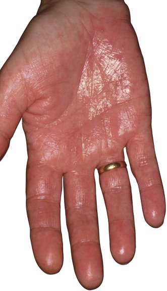
Hyperhidrosis


Hyperhidrosis


James A.A. Langtry


Evidence Levels:  A Double-blind study  B Clinical trial ≥ 20 subjects  C Clinical trial < 20 subjects  D Series ≥ 5 subjects  E Anecdotal case reports


image


Hyperhidrosis is the result of increased secretion of eccrine sweat. This can be annoying, disabling (at work or socially), or indicative of an underlying systemic disease. The eccrine gland is unusual in that the sympathetic sudomotor fibers are cholinergic rather than adrenergic.


Hyperhidrosis can be classified as idiopathic or pathologic.


Idiopathic hyperhidrosis is defined as excessive sweating that is symmetrical, localized to the palms, soles, or axillae (singly or in combination), and independent of thermoregulation. Craniofacial hyperhidrosis is a similar but rarer condition. The other characteristics include its episodic nature, occurrence in response to stimuli, and onset at or after puberty; there is commonly a family history. There is an absence of bromhidrosis and little or no seasonal variation.


Pathologic hyperhidrosis may be localized or generalized. Localized hyperhidrosis may result from injury to the central or peripheral nervous systems, syringomyelia, neuritis, myelitis, tabes dorsalis, or localized vascular diseases, including cold injury, arteriovenous malformation, and erythrocyanosis. Localized hyperhidrosis can occur as a functional nevus in which a normal number of eccrine glands are oversensitive to acetylcholine. Localized areas of hyperhidrosis can develop as a compensatory phenomenon when extensive anhidrosis develops in Ross syndrome (bilateral Holmes Adie pupils, tendon areflexia, generalized anhidrosis). Hyperhidrosis may occur in hereditary conditions, including blue rubber bleb nevus syndrome. The causes of generalized hyperhidrosis include febrile illnesses; metabolic and endocrine diseases (diabetes, hyperthyroidism, gout, acromegaly, pregnancy, porphyria, pheochromocytoma, carcinoid syndrome, alcohol intoxication); congestive cardiac failure and shock; internal malignancy; CNS diseases (tumors and injury); and hereditary syndromes (Chediak–Higashi syndrome and phenylketonuria).


Management strategy


The treatments discussed here apply primarily to the symptomatic management of idiopathic hyperhidrosis.


Local treatments, including medical, electrical, or surgical modalities, aim to stop or reduce sweating sufficiently to control symptoms. Treatments with the lowest risk should be considered first, as dictated by the severity of the condition and in discussion with the patient to assess the balance of risk and benefit.


Topical aluminum chloride hexahydrate (ACH) 2025% solution in ethanol is the first-line treatment. The mechanism of action may result from occlusion of the intraepidermal eccrine duct below the level of the stratum corneum. Correct application technique is critical to compliance: in the axillae, the solution should be applied nightly to the unshaven skin, with or without occlusion, and washed off the following morning before daytime sweating is established. The presence of moisture results in the formation of hydrochloric acid and resultant skin irritation. Mild topical corticosteroids may be used to reduce the common problem of skin irritation, which is the usual reason for treatment failure. ACH solution should not be applied again in the morning. An oral anticholinergic (e.g., 1 mg of sodium glycopyrrolate) 45 minutes before the application of ACH may increase its efficacy by reducing sweating at the time of application, allowing the ACH to be retained on the skin and exert its effect on the sweat pores. The oral anticholinergic may be discontinued after several treatments have initiated a reduction in sweating. Hyperhidrosis may take 2 to 3 weeks to be controlled with topical ACH, at which time application can be reduced to once a week, or at an interval that maintains control.


Other topical therapies include formaldehyde, which is a common contact sensitizer, and glutaraldehyde, which stains the skin. Methenamine gel releases formaldehyde but does not appear to produce contact allergy frequently. The anticholinergic glycopyrrolate in 1.5–2% concentrations topically in an aqueous cream base or as glycopyrrolate pads may be effective.


Iontophoresis is the process of introducing salt ions in solution through the skin into the tissues, and may be effective in treating palmoplantar and axillary hyperhidrosis. Several iontophoretic devices are commercially available. Current is transmitted to electrodes in two trays filled with tap water, and the hands or feet are placed flat in the bottom of the trays. The current is increased until the patient experiences slight discomfort (average 15 mA on the palms and 20 mA on the soles). A special electrode for axillary use is available for some iontophoretic devices. The mechanism of reduction of sweating is not known. Current densities below the threshold of damage to the acrosyringium are employed, and mechanical obstruction does not occur. Iontophoresis is contraindicated in pregnancy and in patients with cardiac pacemakers and metal implants. Twenty-minute sessions three times a week are continued until sweating is sufficiently reduced; thereafter, once- or twice-monthly maintenance therapies are instituted. Anticholinergic drugs such as glycopyrridium bromide may also be introduced by electrophoresis. A recent report suggests that botulinum toxin delivered by iontophoresis to the palms may be effective in the treatment of palmar hyperhidrosis.


Oral anticholinergic drugs and minor tranquilizers produce a dose-related inhibition of sweating and are therefore limited by side effects. Anticholinergic effects, including dry mouth, pupillary dilatation and photophobia, glaucoma, urinary retention, constipation, vomiting, and tachycardia, may, however, occur at doses that produce satisfactory sweat inhibition, thereby limiting their use. The oral anticholinergics most commonly used are glycopyrronium bromide (Robinul), up to 2 mg three times daily, or propantheline 15 mg three times daily. Oral glycopyrronium bromide is not licensed for use in hyperhidrosis in the UK.


Other systemic drugs which have been used include the calcium channel blocker diltiazem, the CNS inhibitor clonidine, and tricyclic antidepressants, although these are the subject of few anecdotal reports.


More aggressive treatments may be sought, as a result of treatment failure, inconvenience of the treatments, or side effects.


Botulinum toxin injected intradermally produces sustained anhidrosis and is used to treat axillary, palmar, facial, and other sites of focal hyperhidrosis. There are eight known serotypes of botulinum toxin, type A exotoxin (BTX-A) being the one that is commercially available as Botox and Dysport. The commercially available BTX-A products differ in their potency and are not equivalent unit for unit. Botulinum toxin type B (BTX-B; Neurobloc) has been shown to be effective in the treatment of axillary hyperhidrosis and may be used as an alternative to BTX-A products, especially where antibody formation has resulted in a loss of clinical benefit to BTX-A. It is recommended that experience be gained with one of the products. BTX-A produces its effect by irreversibly blocking the release of acetylcholine from cholinergic junctions. Treatment of the axilla is simple and well tolerated. Multiple injections, 2 cm apart, are performed in the axillary vault corresponding to the area of maximum sweating (an area of approximately 200 cm2). Palmar skin is painful to inject (may require a regional nerve block), injection is less well tolerated, and there is a potential for producing weakness of the intrinsic hand musculature. It is not a practical treatment for plantar hyperhidrosis. Inactivation of affected cholinergic junctions is permanent, but new cholinergic junctions are produced through the natural process of tissue turnover and repair, so the effect is temporary. Onset of anhidrosis after injection occurs at 24–72 hours and lasts 3 to 6 months. There are reports of efficacy of BTX-A utilizing iontophoresis and dermojet as delivery systems to affected areas.


Surgical and physical techniques that have been employed in the management of axillary hyperhidrosis include:


Selective ablation of the sympathetic innervation of the palms, axillae, and soles reduces sweating effectively. A number of side effects are associated with sympathectomy, including compensatory hyperhidrosis, Horner’s syndrome, pneumothorax, and intraoperative cardiac arrest. Satisfactory long-term results are generally achieved, although recurrence of sweating usually occurs. It is best reserved for severe palmar hyperhidrosis (upper thoracic sympathectomy T2/T3 ganglia), avoiding denervation of the axillary sweat glands and thereby minimizing side effects. This is performed as an open surgical technique or endoscopically (by a transthoracic route) using electrocautery or laser. Percutaneous chemical sympathectomy with ethanol has also been used, a technique that may be employed in lumbar sympathectomy to treat plantar hyperhidrosis. However, this is not generally recommended given the significant risk of sequelae including ejaculatory failure, impotence, and anorgasmia.

Stay updated, free articles. Join our Telegram channel

Aug 7, 2016 | Posted by in Dermatology | Comments Off on Hyperhidrosis

Full access? Get Clinical Tree

Get Clinical Tree app for offline access