Hydroa vacciniforme (HV) and solar urticaria (SU) are uncommon immunologically mediated photodermatoses. HV occurs almost exclusively in children, usually beginning in childhood and remitting spontaneously by adolescence. Association with chronic Epstein-Barr virus infection has been reported in HV, which raises the possibility of lymphoproliferative disorders in these patients. SU is characterized by skin erythema, swelling, and whealing immediately after sun exposure. Although several treatment options are available, the management of both conditions remains a challenge.
Key points
- •
Hydroa vacciniforme (HV) is characterized by a vesiculopapular eruption and necrotic lesions that heal with varioliform scars.
- •
HV has been reported to be associated with latent Epstein-Barr virus infection, raising the possibility of increased risk of lymphoproliferative malignancy.
- •
The mainstay of therapy in HV is adequate photoprotection.
- •
Solar urticaria (SU) is characterized by skin erythema, swelling, and whealing immediately after sun exposure.
- •
Treatments for SU include photoprotection, medical therapy, phototherapy, photochemotherapy, and plasmapheresis.
Hydroa vacciniforme
Hydroa vacciniforme (HV) is a rare photosensitivity disorder predominantly affecting children. It is characterized by recurrent vesiculopapular eruptions that evolve into necrotic crusts and varioliform scars on sun-exposed areas. Latent Epstein-Barr virus (EBV) infection has been suggested to have a role in the underlying pathogenesis. HV has clinically been classified into classic HV and severe HV-like eruption. The latter, described in adults, may be associated with systemic symptoms and an increased risk of lymphoproliferative disorders. Classic HV tends to remit by adolescence or early adulthood. This disease significantly affects quality of life, causing both psychosocial and emotional morbidity.
History
HV was first reported by Bazin in 1862. The term “hydroa” possibly derives from the Greek for “water eggs,” a reference to the vesicular eruption. “Vacciniforme” means “poxlike” scar, which is characteristic of this condition when the lesions heal.
Epidemiology
The rarity of HV and lack of universally diagnostic criteria make the precise prevalence difficult to establish. The estimated prevalence of HV is 0.34 cases per 100,000 population. Although well recognized globally, it predominantly affects Caucasians. A bimodal distribution has been described with peaks presenting at ages 1 to 7 and 12 to 16 years. Several cases of adult-onset classic HV have also been reported. The proportion of female to male patients varies depending on the studies, ranging from 1:1 to 1:2. Male patients tend to have a later onset, longer duration, and more severe symptoms than female patients. Although HV is usually sporadic, familial cases have also been noted.
Pathogenesis
The precise pathophysiology of HV remains unknown. Sunlight, especially ultraviolet A (UVA), is known to play an important role, because the characteristic lesions of HV can be reproduced after artificial UVA exposure. EBV infections may also play a role in the pathogenesis of the disease, as EBV has been detected in the lymphocytic infiltrate of HV lesions from both pediatric and adult patients. Elevated levels of EBV DNA copies in the peripheral blood and EBV-encoded small nuclear ribonucleic acid (EBER) in cutaneous lesions have been found in both classic HV and severe HV-like eruptions. Thus, both conditions have been suggested to be variants within the same disease spectrum. Higher copies of EBV are associated with symptom severity and worse prognosis for patients. In addition to HV, chronic EBV infection is also known to be related to other heterogeneous disorders including lymphoproliferative disorders, hemophagocytic syndrome, and hypersensitivity to mosquito bites. Therefore, patients with the severe variant of HV may be at risk of progression to various EBV-associated malignant lymphomas.
Clinical Manifestations
HV patients experience various clinical signs and symptoms related to their disease. HV is classified into two types: classic or typical HV, and severe HV-like eruption.
Mucocutaneous manifestations
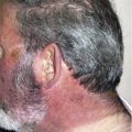
Classic HV usually presents with recurrent erythematous papules and vesicles ( Fig. 1 ) associated with itching or stinging sensation within hours or days after sun exposure. Subsequently, the lesions progress to ulceration with necrotic crusts, and eventually heal over a period of 1 to 6 weeks with varioliform atrophic scars ( Fig. 2 ). The distribution tends to be symmetric. The eruptions typically develop on sun-exposed areas, such as the face and dorsal hands, in the summer. Unlike classic HV, the lesions of severe HV-like eruption are larger and deeper; this variant occurs more commonly in adults. The lesions are distributed extensively, including on sun-protected areas, and may not always be associated with photosensitivity. Facial swelling is also common. Seasonal variation is not observed in the severe form. Disfigurement of the ears and nose, in addition to contracture of the fingers, has been reported. Heat may provoke the symptoms in some of these patients.
Ocular and oral mucosal involvement has been reported in HV. Ophthalmic complications include mild photophobia, chemosis, keratoconjunctivitis, corneal ulcer and erosion, iritis, uveitis, and scleritis ; these were observed in 6.3% of patients in a study from Japan. The same study also reported oral lesions, including aphthous stomatitis and ulcerative gingivitis, in 17.5% of the patients. Oral lesions were seen predominantly in severe HV-like eruptions rather than in classic HV. Classic HV spontaneously resolves during adolescence or young adulthood, whereas severe HV-like eruptions tend to have a relatively longer clinical course and usually become more severe with age.
Systemic manifestations
Classic HV is considered a benign disease with rare complications. However, severe HV-like eruptions may have systemic manifestations, such as fever, malaise, weight loss, hypersensitivity to mosquito bites, lymphadenopathy, hepatomegaly, splenomegaly, abdominal pain, headache, abnormal liver function testing, leukopenia, and thrombocytopenia. Erosions in the esophagus and colon have also been noted. Furthermore, patients with severe disease may progress to EBV-associated malignancy, including T-cell and/or natural killer cell lymphoma.
Histology
Histology of early lesions reveals epidermal spongiosis, focal keratinocyte degeneration, and a perivascular lymphohistiocytic infiltrate. Intraepidermal vesicles, confluent epidermal necrosis, and ulceration are demonstrated in older lesions. Both classic HV and the severe variant share similar histology. However, the severe form usually reveals a denser lymphocytic infiltrate with few atypical cells, which extend into the subcutaneous fat. Immunohistochemical study of the infiltrates usually demonstrates T-cell–expressing cytotoxic molecules, such as T-cell intracellular antigen 1 and granzyme B. Direct immunofluorescence study is usually nonspecific.
Differential Diagnosis
Several conditions need to be considered in the differential diagnosis. Erythropoietic protoporphyria and porphyria cutanea tarda can be differentiated by their characteristic porphyrin profiles. The vesicular form of polymorphous light eruption resolves without significant scarring. Actinic prurigo most commonly occurs in Amerindians or mestizos (individuals with a mixed Amerindian and European extraction). Antinuclear antibody (ANA) panel and skin biopsy should differentiate bullous systemic lupus erythematosus from HV. Herpes simplex virus infection can be diagnosed by viral culture, and contact dermatitis by the history of exposure to contactants.
Laboratory Findings
The diagnosis is generally based on clinical findings and pathognomonic histologic features. In suspicious cases blood, urine, and stool porphyrins may be obtained to exclude cutaneous porphyria. Autoantibody evaluation may be assessed to exclude the low possibility of cutaneous lupus erythematosus. Circulating EBV DNA load may be checked in select cases. Previous reports have proposed that the most useful test to differentiate the two variants of HV is monoclonality of the T-cell receptor genes.
Photobiological Evaluation
Although most patients show increased sensitivity to UVA radiation, they may demonstrate normal minimal erythema dose (MED) to UVA. Repetitive UVA doses have been found to provoke characteristic lesions of HV. The action spectrum for induction of skin lesions ranges from 320 to 390 nm.
Management
An important part of therapeutic management is strict photoprotection, including sun avoidance, use of photoprotective clothing and a wide-brimmed hat, and application of broad-spectrum sunscreen with sun-protection factor higher than 30. Unfortunately, no treatment has been universally successful. Chloroquine, β-carotene, dietary fish oils, prophylactic ultraviolet B (UVB), and psoralen plus UVA (PUVA) have been effective in some cases while thalidomide, azathioprine, and cyclosporine are of uncertain efficacy. In severe cases, systemic corticosteroids can also be used. In cases of chronic EBV infection, antiviral therapy was reported to reduce the severity and frequency of eruptions in a small case series. Because of the potential risk of systemic lymphoma in patients with HV and chronic EBV infection, close monitoring and systemic evaluation should be considered.
The negative impact on the quality of life in HV patients is significant. Psychosocial and emotional impairment in HV patients results from the disfiguring effect of scarring, and the necessity for sun avoidance and consequent restriction of daily activities.
Hydroa vacciniforme
Hydroa vacciniforme (HV) is a rare photosensitivity disorder predominantly affecting children. It is characterized by recurrent vesiculopapular eruptions that evolve into necrotic crusts and varioliform scars on sun-exposed areas. Latent Epstein-Barr virus (EBV) infection has been suggested to have a role in the underlying pathogenesis. HV has clinically been classified into classic HV and severe HV-like eruption. The latter, described in adults, may be associated with systemic symptoms and an increased risk of lymphoproliferative disorders. Classic HV tends to remit by adolescence or early adulthood. This disease significantly affects quality of life, causing both psychosocial and emotional morbidity.
History
HV was first reported by Bazin in 1862. The term “hydroa” possibly derives from the Greek for “water eggs,” a reference to the vesicular eruption. “Vacciniforme” means “poxlike” scar, which is characteristic of this condition when the lesions heal.
Epidemiology
The rarity of HV and lack of universally diagnostic criteria make the precise prevalence difficult to establish. The estimated prevalence of HV is 0.34 cases per 100,000 population. Although well recognized globally, it predominantly affects Caucasians. A bimodal distribution has been described with peaks presenting at ages 1 to 7 and 12 to 16 years. Several cases of adult-onset classic HV have also been reported. The proportion of female to male patients varies depending on the studies, ranging from 1:1 to 1:2. Male patients tend to have a later onset, longer duration, and more severe symptoms than female patients. Although HV is usually sporadic, familial cases have also been noted.
Pathogenesis
The precise pathophysiology of HV remains unknown. Sunlight, especially ultraviolet A (UVA), is known to play an important role, because the characteristic lesions of HV can be reproduced after artificial UVA exposure. EBV infections may also play a role in the pathogenesis of the disease, as EBV has been detected in the lymphocytic infiltrate of HV lesions from both pediatric and adult patients. Elevated levels of EBV DNA copies in the peripheral blood and EBV-encoded small nuclear ribonucleic acid (EBER) in cutaneous lesions have been found in both classic HV and severe HV-like eruptions. Thus, both conditions have been suggested to be variants within the same disease spectrum. Higher copies of EBV are associated with symptom severity and worse prognosis for patients. In addition to HV, chronic EBV infection is also known to be related to other heterogeneous disorders including lymphoproliferative disorders, hemophagocytic syndrome, and hypersensitivity to mosquito bites. Therefore, patients with the severe variant of HV may be at risk of progression to various EBV-associated malignant lymphomas.
Clinical Manifestations
HV patients experience various clinical signs and symptoms related to their disease. HV is classified into two types: classic or typical HV, and severe HV-like eruption.
Mucocutaneous manifestations
Classic HV usually presents with recurrent erythematous papules and vesicles ( Fig. 1 ) associated with itching or stinging sensation within hours or days after sun exposure. Subsequently, the lesions progress to ulceration with necrotic crusts, and eventually heal over a period of 1 to 6 weeks with varioliform atrophic scars ( Fig. 2 ). The distribution tends to be symmetric. The eruptions typically develop on sun-exposed areas, such as the face and dorsal hands, in the summer. Unlike classic HV, the lesions of severe HV-like eruption are larger and deeper; this variant occurs more commonly in adults. The lesions are distributed extensively, including on sun-protected areas, and may not always be associated with photosensitivity. Facial swelling is also common. Seasonal variation is not observed in the severe form. Disfigurement of the ears and nose, in addition to contracture of the fingers, has been reported. Heat may provoke the symptoms in some of these patients.
Ocular and oral mucosal involvement has been reported in HV. Ophthalmic complications include mild photophobia, chemosis, keratoconjunctivitis, corneal ulcer and erosion, iritis, uveitis, and scleritis ; these were observed in 6.3% of patients in a study from Japan. The same study also reported oral lesions, including aphthous stomatitis and ulcerative gingivitis, in 17.5% of the patients. Oral lesions were seen predominantly in severe HV-like eruptions rather than in classic HV. Classic HV spontaneously resolves during adolescence or young adulthood, whereas severe HV-like eruptions tend to have a relatively longer clinical course and usually become more severe with age.
Systemic manifestations
Classic HV is considered a benign disease with rare complications. However, severe HV-like eruptions may have systemic manifestations, such as fever, malaise, weight loss, hypersensitivity to mosquito bites, lymphadenopathy, hepatomegaly, splenomegaly, abdominal pain, headache, abnormal liver function testing, leukopenia, and thrombocytopenia. Erosions in the esophagus and colon have also been noted. Furthermore, patients with severe disease may progress to EBV-associated malignancy, including T-cell and/or natural killer cell lymphoma.
Histology
Histology of early lesions reveals epidermal spongiosis, focal keratinocyte degeneration, and a perivascular lymphohistiocytic infiltrate. Intraepidermal vesicles, confluent epidermal necrosis, and ulceration are demonstrated in older lesions. Both classic HV and the severe variant share similar histology. However, the severe form usually reveals a denser lymphocytic infiltrate with few atypical cells, which extend into the subcutaneous fat. Immunohistochemical study of the infiltrates usually demonstrates T-cell–expressing cytotoxic molecules, such as T-cell intracellular antigen 1 and granzyme B. Direct immunofluorescence study is usually nonspecific.
Differential Diagnosis
Several conditions need to be considered in the differential diagnosis. Erythropoietic protoporphyria and porphyria cutanea tarda can be differentiated by their characteristic porphyrin profiles. The vesicular form of polymorphous light eruption resolves without significant scarring. Actinic prurigo most commonly occurs in Amerindians or mestizos (individuals with a mixed Amerindian and European extraction). Antinuclear antibody (ANA) panel and skin biopsy should differentiate bullous systemic lupus erythematosus from HV. Herpes simplex virus infection can be diagnosed by viral culture, and contact dermatitis by the history of exposure to contactants.
Laboratory Findings
The diagnosis is generally based on clinical findings and pathognomonic histologic features. In suspicious cases blood, urine, and stool porphyrins may be obtained to exclude cutaneous porphyria. Autoantibody evaluation may be assessed to exclude the low possibility of cutaneous lupus erythematosus. Circulating EBV DNA load may be checked in select cases. Previous reports have proposed that the most useful test to differentiate the two variants of HV is monoclonality of the T-cell receptor genes.
Photobiological Evaluation
Although most patients show increased sensitivity to UVA radiation, they may demonstrate normal minimal erythema dose (MED) to UVA. Repetitive UVA doses have been found to provoke characteristic lesions of HV. The action spectrum for induction of skin lesions ranges from 320 to 390 nm.
Management
An important part of therapeutic management is strict photoprotection, including sun avoidance, use of photoprotective clothing and a wide-brimmed hat, and application of broad-spectrum sunscreen with sun-protection factor higher than 30. Unfortunately, no treatment has been universally successful. Chloroquine, β-carotene, dietary fish oils, prophylactic ultraviolet B (UVB), and psoralen plus UVA (PUVA) have been effective in some cases while thalidomide, azathioprine, and cyclosporine are of uncertain efficacy. In severe cases, systemic corticosteroids can also be used. In cases of chronic EBV infection, antiviral therapy was reported to reduce the severity and frequency of eruptions in a small case series. Because of the potential risk of systemic lymphoma in patients with HV and chronic EBV infection, close monitoring and systemic evaluation should be considered.
The negative impact on the quality of life in HV patients is significant. Psychosocial and emotional impairment in HV patients results from the disfiguring effect of scarring, and the necessity for sun avoidance and consequent restriction of daily activities.
Solar urticaria
Solar urticaria (SU) is a rare skin condition characterized by skin erythema, swelling, and whealing immediately after exposure to diverse wavelengths of sunlight. It is estimated that SU accounts for less than 1% of all causes of urticaria. SU most commonly develops in women in the third decade of life. Though uncommon, it has a profound effect on individual quality of life.
History
The term “solar urticaria” was first proposed by Duke in 1923 when he described a 43-year-old woman who developed itching, skin erythema, and edema while she was exposed to the sun during outdoor swimming. In 1923, Wucherpfenning demonstrated that different wavelengths of light could produce urticarial lesions. In 1942, Rajka successfully reproduced urticarial lesions on intradermal injection of an affected patient’s serum into a normal subject. Subsequent information obtained from the serum transfer test helped to clarify the pathogenesis of this condition.
Epidemiology
SU accounts for less than 1% of all causes of urticaria. The prevalence of idiopathic SU in Tayside, Scotland was reported to be 3.1 per 100,000. The frequency of SU in photodermatology referral centers worldwide ranges from 1% to 18%.
Clinical Manifestations
Patients with SU characteristically develop urticarial lesions within 5 to 10 minutes of exposure to the sun; the lesions resolve within 24 hours, usually within 1 to 3 hours. In addition to pruritus, erythema, and edema of the skin, patients may also experience systemic symptoms, such as nausea, dizziness, headache, wheezing, syncope, or, rarely, anaphylactic shock. Whereas most patients develop lesions during direct sun exposure, a minority of patients only develop urticarial lesions immediately or a few minutes after returning to the shade. This latter group was found to have a specific portion of sunlight that acts as an inhibition spectrum. SU commonly affects the upper chest, arms, and forearms, whereas areas that are regularly exposed to the sun, such as the face and hands, are infrequently involved. In highly sensitive patients, SU may occur in covered areas of skin via light penetration through thin clothing.
In addition, there is a rare and less severe form of SU called fixed solar urticaria (FSU). In these patients, the wheals develop exclusively in a fixed area of skin and can be reproduced only at the same site. To date, only 7 patients with this condition have been reported. Recently Wessendorf and colleagues reported a case of delayed-onset FSU, with urticarial wheal development 6 hours after ultraviolet exposure. The pathogenesis of FSU has not been elucidated, but may involve a difference in mast-cell population and distribution in the skin.
Associated Conditions
SU usually occurs in healthy individuals, but has been reported to coexist with other photodermatoses such as polymorphous light eruption, chronic actinic dermatitis, actinic prurigo, and porphyria cutanea tarda. Medications, including benoxaprofen, repirinast, oral contraceptive pills, and tetracycline, have also been reported to induce SU-like reactions.
Differential Diagnosis
The clinical manifestations of SU may resemble other photodermatoses. Detailed history, physical examination, phototesting, and other investigations are essential in arriving at the diagnosis. The onset of eruption within a few minutes of sun exposure and the resolution within a few hours are key in differentiating SU from polymorphous light eruption. Erythropoietic protoporphyria, which may present with urticarial lesions following exposure to sunlight, can be differentiated by its characteristic elevation of protoporphyrin in biological specimens. Systemic lupus erythematosus can be differentiated by ANA panel, and lesions of chronic idiopathic urticaria develop without any correlation with sun exposure. Chronic actinic dermatitis characteristically presents with lichenification on sun-exposed areas. HV heals with scars. Actinic prurigo mainly occurs in Amerindians and mestizos, presenting with persistent papules, cheilitis, and conjunctivitis.
Pathogenesis and Classification
SU results from a type I hypersensitivity reaction. Once skin is exposed to sunlight, a previously inactive cutaneous chromophore is likely converted to a photoallergen. The photoallergen then interacts with immunoglobulin E (IgE)-specific mast cells, causing mast-cell degranulation and clinically apparent wheal and flare.
Studies in the past with passive transfer test, currently considered unethical, have provided significant information on the pathogenesis of SU. This procedure involves intradermal injection of an affected patient’s serum into the skin of a normal subject, followed by irradiation with the causative wavelengths. The reverse passive transfer test involves irradiation of the skin of a healthy individual, followed by injection of serum from the affected patient.
According to Leenutaphong and colleagues, SU is classified into 2 types. In type I, the patient demonstrates an IgE-mediated hypersensitivity to specific photoallergens, which are present only in patients with SU. A patient who has type I SU will have a variably positive passive transfer test and a negative reverse passive transfer test. In type II SU, the patient has abnormal IgE antibodies against a normal chromophore. Patients with type II SU will always have a positive passive transfer test and a variable or negative reverse passive transfer test.
Action Spectrum
The action spectrum in SU refers to the specific wavelength that can produce an urticarial response. The action spectrum of SU varies by geographic and ethnic background, and can range from UVB to infrared wavelength. In a study of 61 patients in France, UVA was the most common action spectrum. In a study of 84 patients in Scotland, 41% of patients reacted to UVA and visible light while 30.9% reacted to visible light alone. A study from Belgium demonstrated that the most common action spectra were UVA alone or UVA and visible light, followed by visible light alone and then UVB alone. In Asians, the most common action spectrum appears to be visible light, followed by a combination of visible light and UVA and UVB. Studies from Japan and Singapore demonstrated that most patients developed urticarial reactions from visible light alone. The infrared spectrum can, in rare instances, precipitate SU; however, in such instances differentiation from heat urticaria could present a challenge because infrared is known to generate heat. The wide range of action spectra may be due to the diversity of photoallergens. Identifying the action spectrum for SU in each patient will allow an individualized treatment plan for photoprotection.
Inhibition Spectrum
Some patients with SU display a delayed urticarial reaction after sun exposure but not while in direct sunlight. This phenomenon can be explained by the theory of inhibition spectrum, which has mainly been reported in the Japanese literature and was first described in 1982 by Hasei and Ichihashi. This landmark study reported a 42-year-old woman who developed urticaria from visible light in the range of 400 to 500 nm, but this reaction was inhibited by immediate re-irradiation with a spectrum longer than 530 nm. In a clinical and photobiological study of 40 patients with SU in Japan, the inhibition spectrum was detected in 68% of patients. In most cases, the wavelength of the inhibition spectrum was longer than that of the action spectrum. The mechanism of the inhibition spectrum is unclear; however, several hypotheses have been proposed, including mast-cell stabilization, photoallergen inactivation, competitive IgE blockade, and blockage between photoallergen and IgE mast cells.
Augmentation Spectrum
After the discovery of the inhibition spectrum, Horio and Fujigaki observed that irradiation with specific wavelengths before irradiation with the action spectrum enhanced the urticarial reaction in a patient with SU. Soon thereafter, Danno and Mori also reported enhanced urticarial response with irradiation of the augmentation spectrum following exposure to the action spectrum, and proposed that the augmentation spectrum may amplify production of photoallergen after irradiation.
Photobiological Evaluation
The purpose of phototesting in patients with SU is to identify the action spectrum of minimal urticarial dose (MUD). Assessment of the phototest sites should be done immediately after irradiation, and ideally up to 1 hour after. The light sources for phototesting should include UVA, broadband UVB, and visible light (usually a slide projector). To eliminate the heat generated by a visible light source, a water filter should be placed between the light source and the irradiated sites. Fig. 3 illustrates an urticarial reaction after visible light exposure. If phototesting results are negative but SU is still suspected, natural sunlight exposure should be considered. In addition, FSU should be considered in patients with recurrent urticarial lesions in a specified area. For FSU, phototesting will only be positive if performed on the previously affected site.