Coinfection with human immunodeficiency virus (HIV) has a major effect on the natural history of many infectious diseases, particularly mycobacterial diseases. Early in the HIV epidemic, it was predicted that HIV infection would worsen leprosy outcomes, with more patients developing lepromatous disease, an impaired response to multidrug therapy and fewer reactions. However, studies on the epidemiologic and clinical aspects of leprosy suggest that the course of leprosy in coinfected patients has not been greatly altered by HIV. In contrast, initiation of antiretroviral treatment has been reported to be associated with activation of subclinical Mycobacterium leprae infection and exacerbation of existing leprosy lesions. With regular new discoveries about the interaction of leprosy and HIV, the need to maintain research in this field is of considerable importance.
Co-infection with HIV has a major effect on the natural history of many diseases, particularly mycobacterial diseases. Early in the HIV epidemic it was predicted that HIV infections would worsen outcomes in leprosy patients with more patients developing lepromatous disease and patients having fewer immune reactions. Now that many patients receive HAART tuberculoid leprosy types predominate and reactions are an important clinical feature in co-infected patients.
Leprosy is a chronic infectious disease affecting nerves and skin. It has a long incubation period of 2 to 10 years, and presents with a clinical spectrum depending on the relationship between the host immune system and the bacteria. At one end of the spectrum is tuberculoid leprosy, characterized by strong cell-mediated immunity (CMI) toward M leprae . These patients have few hypopigmented, anesthetic lesions. At the other pole is lepromatous leprosy (LL), which is characterized by the absence of a CMI response. These patients have numerous lesions and high bacillary loads. Most patients have features between these two extreme groups and fall in the categories of borderline tuberculous (BT), borderline borderline (BB) or borderline lepromatous (BL). The borderline cases are immunologically unstable and at greater risk of type 1 reaction, which affects mainly the nerves and skin. The lepromatous types of BL and LL are at higher risk of erythema nodosu leprosum (ENL), a more systemic and severe immunologic complication.
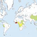
In 2008, 121 countries reported a total of 249,007 new leprosy cases to the World Health Organization (WHO). Most endemic countries for leprosy also have a high HIV prevalence, increasing the possibility of HIV–leprosy coinfection.
The few published small studies provide limited data on the course of leprosy in coinfected patients. HIV incidence was not found to be increased among leprosy patients compared with nonleprosy groups. All types of leprosy can occur in coinfected patients. Two East African studies reporting an increase multibacillary (MB) cases. However since the introduction of HAART borderline tuberculoid leprosy is the predominant form, as reported in Brazilian studies. Coinfected patients treated with standard length WHO-multi-drug therapy (MDT), have responded adequately, although there might be a possibility of an increased relapse rate. A Ugandan study demonstrated an increased risk of developing type 1 reactions in an MB leprosy patient with HIV, and increased recurrence rates of type 1 reactions were seen in an Ethiopian study. In general, however, neuritis was not found to be more severe in HIV-positive cases. A few case reports of ENL in coinfected patients have been published. Co-infected patients with reactions appear to need very long courses of steroid treatment. Patients with HIV are also at risk of developing peripheral nerve damage including generalized peripheral neuropathy and mono-neuritis multiplex through several mechanisms, namely, treatment with antiretrovirals and HIV infection per se. In analogy to the situation for tuberculosis in HIV coinfected individuals, it was assumed that HIV coinfection would worsen nerve damage in leprosy patients. There are a few early studies reporting no increase in nerve damage in coinfected patients. A well controlled study of peripheral nerve function in coinfected patients would be useful. Table 1 summarizes the expected versus actual impact of HIV-1 on coinfected patients.
| Theory | In Practice | ||
|---|---|---|---|
| Epidemiologic | Incidence | Increase in leprosy | No change |
| Clinical | Tuberculoid leprosy | Decreased | Increased |
| Treatment response | Worsened | No change | |
| Type-1 reactional states | Fewer | Increased | |
| Neuritis | Worsened | ? | |
| Novel findings | Presentation as immune reconstitution inflammatory syndrome | ||
| Histopathological | Granuloma formation | Decreased | No change |
| Bacterial index | Increased | No change | |
Immunology of HIV and leprosy coinfection
Patients with tuberculoid leprosy have good cell-mediated immune response to M leprae, resulting in a few skin lesions, which histologically have well organized lymphocyte (CD68+, CD3+, CD8+, CD4+)-rich granulomas with predominantly CD4 T cells. In contrast, patients with LL have a strong humoral response but poor or absent cell-medicated immunity, resulting in uncontrolled growth of bacilli and disseminated skin lesions. Histologic examination of biopsies from their lesions reveals that the granulomas are comprised of macrophages and small numbers of CD8 T cells.
HIV affects cell-mediated immunity, and it was initially expected that, just as in M. tuberculosis infection, the decrease in CD4 cells would result in decreased capacity for mycobacterial containment and thus an increase in disseminated disease. But studies have shown that HIV coinfected patients with low CD4 count had borderline tuberculoid lesions with well formed granuloma and normal CD4 cells numbers. In contrast, coinfected patients with LL lesions showed loose infiltrates comprised of macrophages and a small number of almost exclusively CD8 lymphocytes. Carvalho and colleagues found that the coinfected group exhibits lower CD4 to CD8 ratios, higher levels of CD8+ activation, increased Vδ1 to Vδ2 T cell ratios and decreased percentages of plasmacytoid dendritic cells as compared with HIV-1 mono-infected patients. The exact immunopathological mechanism underlying the possible increase in frequency of leprosy reactions is not clear. Dysregulation of the immune system and the heightened state of immune activation in HIV infection may be responsible. In addition, delayed clearance of M.leprae antigen caused by impaired phagocytic function of macrophages also has been implicated.
Effect of antiretroviral therapy on HIV and leprosy coinfection
Since the introduction of highly active antiretroviral therapy (HAART) in the management of HIV, especially in regions endemic for leprosy, co-infected patients are also developing tuberculoid leprosy with active lesions, and ulceration of lesions is seen in leprosy type 1 reactions. Leprosy is being increasingly reported as part of the immune reconstitution inflammatory syndrome (IRIS).
IRIS is a paradoxic deterioration in clinical status after starting HAART, a deterioration that is attributable to the recovery or reactivation of someone’s immune response to a latent or subclinical process. The HAART regimes currently used increase production and redistribution of CD4+ cells and improve pathogen-specific immunity, both to HIV and other pathogens. While improved immunity to HIV is the required effect from HAART, improved immunity to other opportunistic pathogens or development of autoimmunity can result in IRIS. The prevalence of IRIS in cohort studies of HIV-positive patients ranges from 3% to more than 50%, varying greatly with the acquired immunodeficiency syndrome (AIDS)- defining illness affecting the patient at the start of HAART therapy. Risk factors for the development of IRIS include advanced HIV disease with a CD4+ T cell count under 50 cells/mm, unrecognized opportunistic infection or high microbial burden, and the number and presence of prior opportunistic infections. HAART triggers overt clinical manifestations of coinfection with tuberculosis, cytomegalovirus, herpes zoster, C and B hepatitis virus, and now leprosy.
Since 2003, 23 reports of patients developing leprosy as IRIS have been published. Most patients had borderline leprosy, and type 1 reactions frequently occurred. Ulceration, a highly unusual feature in leprosy lesions, was observed in six patients, and four patients also developed neuritis. It may be that the high proportion of borderline tuberculoid leprosy cases among the HIV-infected patients on HAART could shed light on the questions related to the kinetics of M leprae infection and development of disease. Borderline tuberculoid leprosy manifests 2 to 5 years after infection, at which time specific cell-mediated immunity is strong enough to cause tissue damage as well as kill or at least control mycobacterial growth. On the other hand, lepromatous (BL and LL) forms appear in patients after longer periods of incubation (5 to 10 years), during which time a large number of bacilli have accumulated in the tissue due to progressive reduction of CMI. Although HIV patients are typically more aware of their health status and would easily detect small lesions, the high frequency of borderline tuberculoid patients and reactions among HIV-infected individuals and the low bacillary load among the co-infected MB patients strongly suggest an earlier-than-usual detection of the disease in immune-reconstituted patients.
To facilitate the recognition and classification of leprosy-associated IRIS, the following case definition has been suggested :
Leprosy or leprosy reaction presenting within 6 months of starting HAART
Advanced HIV infection
Low CD4+ count before starting HAART
CD4+ count increasing after HAART has been started.
Subdividing leprosy-associated IRIS into groups according to data on timing and clinical presentation may help toward defining the causes and mechanisms of this phenomenon. Deps and Lockwood recently proposed such a subdivision, attempting to separate unmasking episodes from those of overlap of immune restoration.
There are several possible mechanisms for the pathogenesis of leprosy IRIS. Leprosy has a long incubation period, and HAART may provide the immunologic trigger for normal disease. Another explanation is that leprosy-associated IRIS is similar to a type 1 reaction. Whatever the underlying mechanisms, it is likely that leprosy-associated IRIS will be increasingly reported, especially as access to HAART becomes more widely available. There are also several case reports of patients being diagnosed with leprosy more than 6 months after the initiation of HAART. Although these patients can present with any kind of leprosy, including histoid leprosy, the time lag of greater than 6 months since the initiation of HAART excludes the diagnosis of IRIS.
Related posts:
 Buruli Ulcer: Advances in Understanding Mycobacterium ulceransInfection
 Outbreak of Nontuberculous Mycobacterial Disease in the Central Pacific
 Arsenical Keratoses in Bangladesh—Update and Prevention Strategies
 Chagas Disease: Coming to a Place Near You
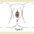 Female Genital Mutilation: What Every American Dermatologist Needs to Know
 Widespread Use of Toxic Skin Lightening Compounds: Medical and Psychosocial Aspects
Stay updated, free articles. Join our Telegram channel

Full access? Get Clinical Tree


