Fig. 1.1
Wilhelm Conrad Röntgen with his wife Anna Bertha Ludwig and the coachman Emanuel Schmid who used to drive them regularly up to the Engadin in the Swiss Alps for summer vacation (Archive of the Institute for the History of Medicine, University of Zurich)
On the evening of November 8th 1895 Wilhelm Conrad Röntgen, at that time professor of physics in Würzburg, discovered a “new kind of rays”, as he published in January 1896 in the “Sitzungsberichte der Würzburger Physikalisch-medizinischen Gesellschaft” [1].
The news about these miraculous rays spread very rapidly all over the world. At the same time, as Röntgens’ article was published in Nature and Science, the fascinated public was already able to admire this curiosity in public demonstrations, for example, in a theater in Davos [2].
Immediately, many researches began to study x-rays, and the biologic effects of radiation became quickly apparent through signs of damage of the skin. Radiation-induced dermatitis was reported in March 1896 and depilation and pigmentation in April 1896 [3].
These reports led Leopold Freund, Dermatologist in Vienna, to use x-rays on a pigmented hairy nevus in November 1896. The treatment resulted in epilation and after 2 months in an ulcer which rapidly became deep and painful and ultimately gave rise to a carcinoma with metastases [4].
Freund described his experiences in 1903 in the book Grundriss der gesammten Radiotherapie for the practitioner [4]. After Freund’s publication, x-rays were tested empirically on almost all skin affections. Among the very early indications for x-ray treatment was the treatment of fungal infections of the scalp, mainly favus and microsporia. Radiotherapy became the gold standard for the treatment of such indications up to 1958 when griseofulvin came on the market [5] (Figs. 1.2, 1.3, 1.4, 1.5, 1.6, 1.7, and 1.8).
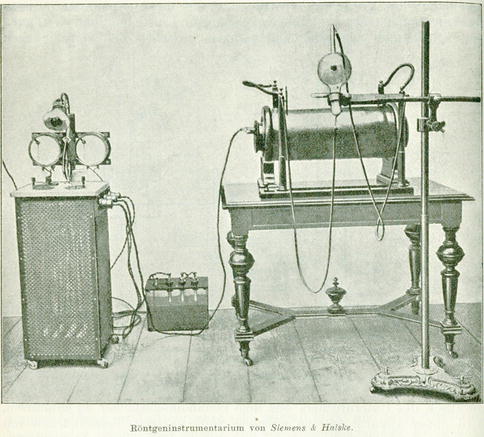
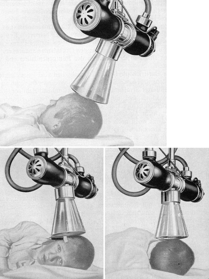

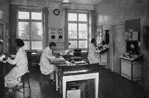
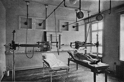
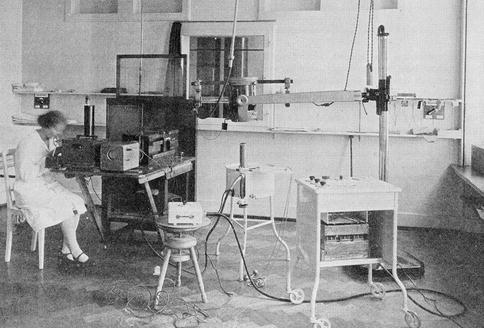
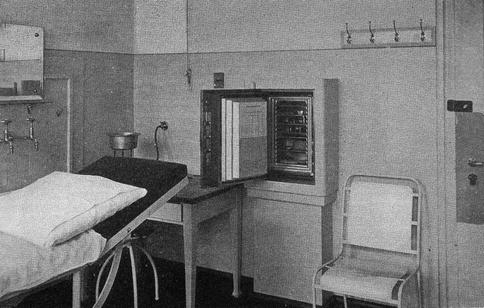
Fig. 1.2
“Röntgeninstrumentarium” (Freund L (1903) Grundriss der gesammten Radiotherapie, Urban & Schwarzenberg, Berlin Wien)
Fig. 1.3
Depilation treatment of trichophytia of the scalp (Blumenthal F, Böhmer L (1923) Strahlenbehandlung bei Hautkrankheiten. Karger, Berlin 1932)
Fig. 1.4
Moulage No. 207: Radiodepilated scalp with microsporia. Made in 1918 by Lotte Volger, Dermatology Clinic Zurich (Museum of Wax Moulages, University and University Hospital Zurich)
Fig. 1.5
Controlling room for radiotherapy at the clinic in Zurich in 1926 (Bloch B (1929) Die Dermatologische Universitätsklinik Zürich. Methods and Problems of Medical Education, The Rockefeller Foundation, New York)
Fig. 1.6
Treating room for radiotherapy at the clinic in Zurich in 1926 (Bloch B (1929) Die Dermatologische Universitätsklinik Zürich. Methods and Problems of Medical Education, The Rockefeller Foundation, New York)
Fig. 1.7
Room for measuring Rx radiation at the clinic in Zurich in 1926 (Bloch B (1929) Die Dermatologische Universitätsklinik Zürich. Methods and Problems of Medical Education, The Rockefeller Foundation, New York)
Fig. 1.8
Safe for the storage of radium at the clinic in Zurich in 1926 (Bloch B (1929) Die Dermatologische Universitätsklinik Zürich. Methods and Problems of Medical Education, The Rockefeller Foundation, New York)
1.2 Indications for X-Ray Treatment
The best results were achieved in the treatment of any kind of eczema and psoriasis. Children with mycosis of the scalp and with port-wine stains were treated successfully with x-rays. Tuberculosis of the skin, also treated with the Finsen UV light, seemed to respond in most cases. But there were also warnings about the possible risk of developing lupus vulgaris carcinoma.
X-ray treatments against acne and rosacea did not work well. Case reports have been published of patients having been treated with x-rays of different quality and quantity for almost a year without improvement.
There was a debate about whether cancer of the skin should be treated with radiation. In 1899, Thor Stenbeck in Stockholm treated a patient with skin cancer of the nose with success when applying small doses of Röntgen rays in daily sessions over a period of several months [6]. On some types of epithelioma, what we call basal cell carcinoma today, x-rays seemed to work very well, while others were refractory [7].
One of the pioneering publishers on the subject of good outcomes in skin cancer treatment with x-rays was the dermatologist Guido Miescher. Together with Bruno Bloch, he had come from Basel to Zurich when the clinic was founded in 1916 and followed Bloch in 1933 as Director of the Clinic and ordinary professor for dermatology in Zurich (Fig. 1.9).
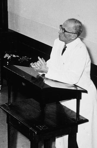
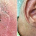
Fig. 1.9
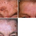
Guido Miescher giving a lecture: it is recognizable that Miescher had acquired a chronic radiodermatitis on the cheeks and the chin. It is verbally passed on that he had himself radioepilated either because he wanted to avoid arduous daily shaving or for medical reasons like a folliculitis (Department of Dermatology, University Hospital Zurich)
Related posts:

Stay updated, free articles. Join our Telegram channel

Full access? Get Clinical Tree


