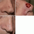
Fig. 8.1
Breadloaf technique. (a) Standard excision processed with breadloaf technique (Image ©2016, Memorial Sloan Kettering Cancer Center. Used with permission). (b) Melanoma excised with standard margin as an ellipse. (c) Melanoma specimen processed with breadloaf technique. (d) Sampling error: red asterisk shows missed tumor at peripheral margin with breadloaf technique (Image by Kishwer S. Nehal)
Advantages
The breadloaf technique has many advantages compared to other processing methods, highlighting its popularity for standard excisions [25]. Small skin specimens bode fairly well and are easy to interpret in this manner. More importantly, there is a clear distinction between central tumor and peripheral margin, with the ability to detect a narrow margin of clearance. This technique allows for better assessment of the field of damage, without the need for a “normal control” specimen for clarification.
Limitations
However, the breadloaf technique is not without limitations. More sections are often required for better examination of margins. The major limitation is lack of full margin assessment, with often less than 1 % of the total peripheral and deep margin microscopically examined. This can lead to the potential for missed tumor at margins between evaluated sections (Fig. 8.1d) [26]. In LM, this is particularly worrisome as some authors feel that the tumor extends in subclinical projections that may be easily missed in the discarded portions between sections [28, 29]. In one recent study, patients with LM were at a greater risk of persistent disease in wide local excisions compared to other subtypes of melanoma, despite a reported negative margin on excisional biopsy [30]. Immediate reconstruction of surgical defects with complex flaps or grafts without confirmation of clear surgical margins further complicates the situation as it is often difficult to pinpoint location of residual melanoma following tissue rearrangement. Thus excision with standard surgical margins is often ineffective in LM/LMM prompting the need for methods with more complete margin control [5, 8, 9, 11–17, 23].
Margin Controlled Excision Techniques
The focus on LM treatment has more recently shifted toward techniques with more meticulous margin evaluation, including en face and radial sectioning and processing with frozen or permanent sections. Each of these techniques has the advantage of more complete margin evaluation and low local recurrence rates of 0–5 % [31–37] compared to standard excision with local recurrence rates of 8.8–20 % [3, 5, 23]. Unique, distinguishing characteristics of each margin controlled technique will be reviewed.
Mohs Micrographic Surgery Using Frozen Sections
Technique
A modified en face sectioning with frozen sections is used in MMS [10, 14, 15, 18, 20–22, 31, 37] and offers the advantage of complete margin control. The MMS technique relies on two main principles in order to achieve success in tumor clearance: (1) contiguous tumor growth for microscopic mapping, and (2) accurate histologic assessment of tumor cells and differentiation from non-tumor cells on frozen sections [37]. MMS for melanoma involves clinically demarcating the central pigmented lesion with a margin of normal-appearing tissue. This central tumor debulking specimen is excised and sent for serial sectioning [14] with permanent histology for confirmation of tumor depth and staging information. An additional margin is excised to the subcutaneous plane and the complete peripheral and deep margins are processed according to the Mohs technique into frozen sections and evaluated by the Mohs surgeon [14] (Fig. 8.2). The Mohs surgeon microscopically maps residual tumor which guides subsequent Mohs excisions until a tumor-free plane is obtained.
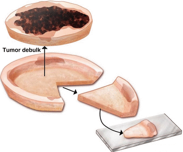
Fig. 8.2
Mohs micrographic surgery technique: tissue is processed with frozen sections for complete margin evaluation (Image ©2016, Memorial Sloan Kettering Cancer Center. Used with permission)
Advantages
The MMS frozen-section technique offers several advantages over standard excision for LMM: (1) tumor with poorly defined clinical margins and unpredictable subclinical extension can be mapped with virtually 100 % margin control; (2) maximal preservation of normal tissue in the anatomically and cosmetically sensitive region of the head and neck, (3) immediate tissue processing and evaluation with same-day repair minimizing wound care and transportation burdens for the patient and (4) low local recurrence rates compared to standard excision [13, 14, 21, 37–42].
Limitations
Most criticism posed against MMS for melanoma involves the quality of frozen sections versus the gold-standard permanent paraffin-embedded sections . Processing high quality thin frozen sections for LM margin evaluation while avoiding tissue distortion and freeze artifact requires highly skilled histotechnicians [41]. Additionally, melanocytes on frozen sections lose their characteristic retraction halo seen on permanent sections, making identification more challenging (Fig. 8.3) [14, 37]. Other pitfalls on Mohs frozen sections include presence of inflammation obscuring tumor, severe keratinocytic atypia within epidermis, and identifying solitary isolated atypical melanocytes [14, 37, 41]. Single melanocyte spread is particularly challenging to identify, as this often occurs in chronically sun damaged skin and distinction with frozen sections can be difficult [37].
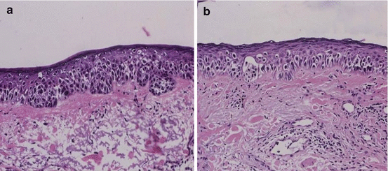
Fig. 8.3
Frozen vs. permanent section evaluation of lentigo maligna. (a) Frozen section (H&E). (b) Paraffin-embedded permanent section (H&E)
From a quality assurance perspective, another disadvantage of MMS for melanoma margin assessment is the lack of a routine independent review of every case by another pathologist (as is routine for intraoperative frozen sections). While MMS may achieve negative margins with low recurrence rates there is lack of independent verification whether all the layers during a procedure that were taken were truly necessary (some may have been falsely read as positive leading to greater tissue defects than medically required).
One alternative to overcome the pitfalls of frozen section evaluation of LMM margins is to excise tissue with the Mohs technique but send the specimen for paraffin embedded en face sectioning. Another option is to excise an additional layer and send for permanent sections at the conclusion of MMS with final margin confirmation by a dermatopathologist [13, 18, 40]. Most studies using this method of checks and balances have found reasonable correlation between frozen and permanent sections [40, 42], however one small study demonstrated higher recurrence rates in frozen MMS sectioning (33 %) compared to rush permanent serial sectioning (7.3 %) in a follow-up period approaching a mean of 10 years [39]. To address challenges of interpreting LMM margins on Mohs frozen section with hematoxilyn & eosin (H&E) staining alone, melanocytic immunostains are commonly used as outlined in the Mohs Surgery chapter.
Staged Excision with Rush Permanent Sections with En Face Sectioning
Technique
En face sectioning of LMM margins can also be assessed with paraffin embedded rush permanent sections to ensure complete margin control while avoiding frozen section pitfalls and need for immunostains. The tumor is clinically demarcated and removed with a surrounding margin of normal appearing tissue ranging from 2 to 10 mm based on the anatomic location and depth of melanoma invasion identified on biopsy [9, 28, 38, 43, 44]. The central portion of the tumor is debulked and processed with serial sectioning or traditional breadloaf sectioning for identification of unsuspected invasion and determination of final Breslow depth for melanoma staging. The perimeter is then divided into smaller sections, often inked for orientation, and the outer-facing rim (true surgical margin) is mounted flat, serially sectioned vertically, and stained with hematoxylin and eosin for examination by a dermatopathologist. This allows for complete peripheral margin evaluation with precise mapping of residual tumor (Fig. 8.4 ). Generally speaking a geometric, sharp angled border is easier to section vertically [32, 44], however despite the change in the shape of the strips, the contoured technique is not reported to have processing difficulties [34]. If a positive margin results, the process is repeated in the mapped area until tumor free margins are achieved. The exception is the square technique, which leaves the central tumor intact until the peripheral margins are clear [32]. The remaining central tumor is then removed and processed in the final steps.
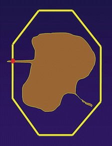
Fig. 8.4
Staged excision with en face permanent sections: Red dot shows tumor detected at peripheral margin (Image by Kishwer S. Nehal)
Advantages
While technically tedious for histotechnicians, the en face technique allows for fewer sections to be examined by the dermatopathologist when compared to the traditional breadloaf method. Most importantly, full evaluation of margins allows for the removal of significantly smaller margins in successive steps until tumor-free margins are achieved, thereby minimizing the cosmetic defect while reducing the rates of local recurrence [9, 28]. One study directly compared the local recurrences rates of conventional excision versus en face sectioning with rush permanent sections and found a 0.7 % local recurrence rate in the en face group compared to 6.4 % with conventional excision [28]. Furthermore, these authors found a significant effect of en face sectioning on recurrence-free survival in patients with thinner LMM (less than or equal to 1 mm in depth). Another study showed no local recurrence using en face techniques, although duration of follow up was not specified [43].
Limitations
The main disadvantages of the en face sectioning technique include inability to define the margin of tumor clearance, and difficulty in tracking out the subtle changes of the trailing edge of LM, with less precise distinction between lesion and background. Some have suggested taking a sample of “normal” yet equally sun-damaged skin to act as a control specimen. However this results in an additional wound/scar for the patient, which is not ideal [9, 45]. Additionally, sections tend to be large and tedious to process, with difficulty obtaining quality sections [25]. Formalin fixation may warp tissue, reduce tissue pliability, and pose a challenge to obtaining a complete en face peripheral margin (Fig. 8.5). However, one group proposed a variation to the traditional fixation technique to reduce this complication by gently fixing the tissue specimen between two glass slides to maintain the flat margin architecture during formalin processing [46]. Furthermore, en face sectioning relies entirely on a contiguous tumor growth pattern, and does not account for possible skip areas within the tumor itself, potentially complicating perfect margin control.
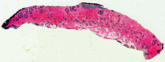
Fig. 8.5
Technical challenge with en face processing: incomplete epidermal margin
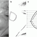
Staged Excision with Rush Permanent Sections with Radial Sectioning
Technique
Radial sectioning is an alternative method for processing LMM margins. Staged excision with rush paraffin-embedded permanent sections using radial sectioning offers the advantage of enhanced margin examination along with the ability to view the transition from tumor to background photodamaged skin (Fig. 8.6a) [8, 17]. The clinical pigmented lesion is demarcated with the use of a Wood’s lamp, and a margin of normal-appearing skin is marked based on initial biopsy characteristics (5 mm margin for melanoma in situ, 7 mm margin for melanoma in situ with regression or radial growth phase microinvasion, and 10 mm margins for invasive melanoma ≤1 mm in Breslow depth) (Fig. 8.6b). The central pigmented lesion or tumor debulking is excised to the deep subcutis and sent to pathology for serial vertical sections to determine final melanoma depth and pathologic staging. The peripheral margins are excised separately to the deep subcutis, divided into 4 quadrants, similar to the face of the clock (12–3, 3–6, 6–9, 9–12 o’clock with sutures to maintain orientation) placed in separate formalin containers (Fig. 8.6c), and sent for rush paraffin-embedded permanent sections. The peripheral margins are inked to identify the inner and the true outer surgical margin, sectioned radially at 1–2 mm intervals in clockwise orientation, and each section placed in a separate cassette (Fig. 8.6d) for processing. Tissue is stained with hematoxylin and eosin, and evaluated by a dermatopathologist and location of residual melanoma communicated to the surgeon. Further excisions are performed based on mapped location of residual LM until margins are histologically cleared. Clear margins are defined as a 3 mm distance between LM and the nearest side margin where anatomically feasible. Studies with 5 years of followup show a 5 % local recurrence rate [17, 47, 48].
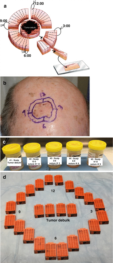
Fig. 8.6
Staged excision with radial sectioning technique. (a) Illustration shows tumor debulking and margins excised (Image ©2016, Memorial Sloan Kettering Cancer Center. Used with permission). (b) Melanoma in situ marked with 5 mm excision margin. (c) Tumor debulking and peripheral margins placed in separate formalin containers. (d) The divided tissue is placed into cassettes for processing and embedding with complete preservation of tissue orientation
Advantages
Advantages of the radial sectioning technique include the ability to evaluate tumor margins compared to central tumor and determine margin of clearance, unlike the en face technique. In this technique, the radial or centrifugal orientation of the specimen shows a clear transition between LM/LMM, atypical junctional hyperplasia, and normal histologic features. This enables detection of subtle changes in cellular density from the central melanoma to the periphery (Fig. 8.7a), which serves as a critical factor when distinguishing tumor-involved margin from chronically sun-damaged background changes. For this reason, immunostains and “normal” control biopsies are rarely needed for a definitive diagnosis/margin evaluation as there is an inherent control when examining the specimen in a radial fashion. The radial evaluation of LMM and its margins also allows the dermatopathologist to measure a margin of clearance (Fig. 8.7b). Our experience suggests that narrow margins of clearance can increase risk of local recurrence over time.
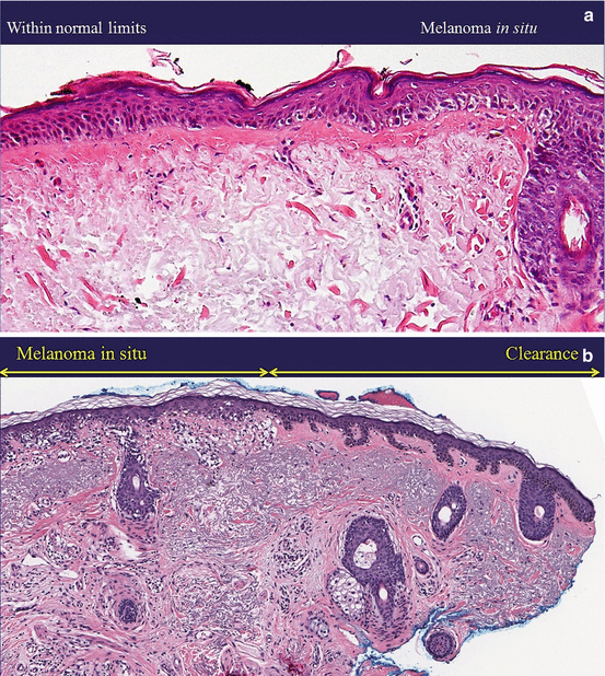

Fig. 8.7
Radial sectioning advantages. (a) Clear visualization of melanoma in situ centrally as it transitions to normal sun damaged skin at surgical margins peripherally (H&E). (b) A margin of clearance can be measured from the outer surgical margin inked in blue (H&E). (c) Tumor can still be detected at peripheral margins (red dots) (Image by Kishwer S. Nehal)
Limitations
Although serial vertical sections are more easily processed in a general pathology laboratory that may be unfamiliar with Mohs or en face section techniques, this technique does require more total slides to be examined by the dermatopathologist which can be time consuming. In addition, this method cannot examine 100 % of the peripheral margin compared to the en face technique or MMS. However, it is unlikely that LM at a peripheral margin would be missed with this technique that examines multiple thin sections (2 mm) and uses a 3 mm safety margin of clearance (Fig. 8.7c). Reconstruction is not same-day, and the overall process may be time-consuming with 24-h tissue processing turn-around time between serial excisions. As with any technique, intimate coordination of care and frequent communication between oncologic surgeon, pathologist and plastic surgeon is essential.
Immunohistochemistry
Immunohistochemistry (IHC) for melanocyte differentiation antigens may be used for the diagnosis of melanocytic neoplasms on both permanent and frozen sections. Markers to visualize intraepidermal melanocytes include MART-1/Melan-A, Sox10, Tyrosinase, Mel-5, and MITF (Fig. 8.8). S100 protein and HMB-45 are less suitable due to limitations in sensitivity (HMB-45, S100P) and specificity (S100P). Although rapid and efficient processing systems have been developed [49–51], they still require an additional 20–40 min on average per section [52]. Additionally, the process is highly technical and as previously stated, requires a skilled and experienced laboratory [1].
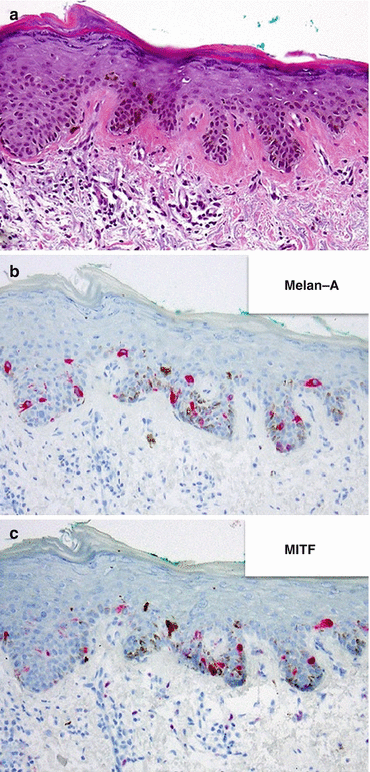
Fig. 8.8
Immunohistochemistry for melanocyte density. (a) H&E. (b) Melan—A. (c) MITF
Melanoma recognized by T cell antigen 1 (Melan-A, MART-1) staining is more commonly used due to its high sensitivity and specificity [15, 53]. It is a cytoplasmic melanosome-associated glycoprotein that stains adult melanocytes, melanomas, and nevus cells [38, 54]. Microphthalmic transcription factor (MITF) and SOX10 are nuclear antigens, which is beneficial for analyzing heavily pigmented lesions [38, 55]. For the recognition of invasive desmoplastic melanoma, S100 protein Sox10 and NFGR are the best markers.
IHC is rarely necessary for a high quality formalin-fixed and paraffin-embedded hamtoxylin and eosin-stained section [8]. IHC may be needed, if the melanocytes are difficult to see due to poor staining or when a dense inflammatory cell infiltrate obscurese the junctional melanocytes (Fig. 8.9). Frozen and en face sections more heavily rely on immunostains for assessing melanocyte density and growth patterns . Interestingly, in one survey from 2000, less than 15 % of MMS laboratories were using immunostains in LM [56]. However, with improved technologies and staining processes and more operator comfort with stains, it is uncertain if this figure is now higher.
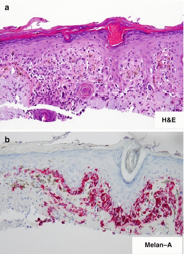
Fig. 8.9
Immunostains for Lentigo Maligna. (a) Inflammation can obscure a junctional melanocytic proliferation (H&E). (b) Melan-A highlights the melanocytic proliferation
Pathologic Staging and Lymph Node Management
Once the LM and LMM has been completely excised, pathologic staging can be completed according to American Joint Committee on Cancer [57] as with other subtypes of melanoma. The recommendations for management and workup of invasive melanoma are outlined in National Comprehensive Cancer Network clinical practice guidelines [58].
Tumor staging is determined by Breslow depth and presence or absence of ulceration and/or mitoses (57). In situ melanoma (LM) is stage Tis. T1 tumors have a thickness ≤ 1.0 mm, with T1a tumors showing no ulceration and mitosis <1/mm2, and T1b tumors with ulceration or mitosis ≥ 1/mm2. T2 tumors have a thickness of 1.01–2.0 mm, with T2a tumors without, and T2b with ulceration. T3 tumors have a thickness of 2.01–4.0 mm, with T3a without, and T3b with ulceration. T4 tumors are >4.0 mm in thickness, with T4a without, and T4b with ulceration.
Nodal staging is determined as follows: NX patients are those in whom the regional lymph nodes cannot be assessed (57). N0 patients have no regional metastasis. N1a category refers to those with 1 node containing micrometastasis (found on sentinel lymph node biopsy or lymph node dissection), while N1b refers to 1 node containing macrometastasis (clinically detectable). N2 category refers to patients with 2–3 nodes classified by the following subcategories: a. micrometastasis, b. macrometastasis, or c. In transit met/satellite without metastatic nodes. N3 category includes those with 4 or more metastatic nodes, matted nodes, or in transit mets/satellites with metastatic nodes. Distant metastasis M categories are determined as follows: M0 is no detectable evidence of distant metastases; M1a includes metastases to the skin, subcutaneous tissue, or distant lymph nodes; M1b includes metastatic disease to the lung; M1c includes metastases to all other sites or distant metastases in combination with increased serum LDH levels.
Related posts:


Stay updated, free articles. Join our Telegram channel

Full access? Get Clinical Tree


