Graft-Versus-Host Disease

44


Graft-Versus-Host Disease


Multiorgan disorder that most commonly results from the transfer of donor hematopoietic stem cells into a recipient via an allogeneic hematopoietic stem cell transplant (HSCT).


Despite advances in HSCT procedures and post-transplantation immunosuppressive therapy, more than half of HSCT recipients develop chronic graft-versus-host disease (GVHD), which remains a major cause of morbidity and mortality.


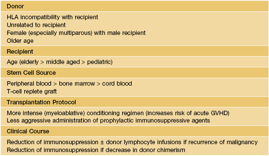
Table 44.1 lists risk factors for GVHD in HSCT recipients.


Less frequent settings of GVHD include transfusion of non-irradiated blood products to an immunocompromised patient, maternal–fetal transmission to an immunodeficient neonate, and solid organ transplantation.


Divided into acute and chronic forms based on clinical features.


Pathogenesis of acute GVHD occurs in three steps: (1) HSCT conditioning regimen leads to epithelial cell injury and activation of host antigen presenting cells; (2) activation of donor T cells; (3) tissue destruction by cytotoxic T cells, natural killer cells, and soluble factors (e.g. tumor necrosis factor-α [TNF-α]).


Chronic GVHD shares features (e.g. autoantibody production, cutaneous sclerosis) with autoimmune connective tissue diseases.


Histologically, acute GVHD and epidermal involvement in chronic GVHD are characterized by variable degrees of keratinocyte necrosis (often accentuated in appendages), vacuolar degeneration of the basal layer, and a band-like lymphocytic infiltrate.


Acute GVHD


Most often arises 4–6 weeks after HSCT with traditional regimens.


Persistent, recurrent, and late-onset variants of acute GVHD can occur during a time period (>100 days post-transplant) traditionally reserved for chronic GVHD, especially following interventions such as tapering of immunosuppression and donor lymphocyte infusions.


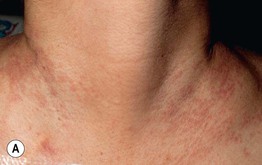
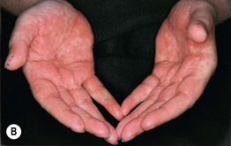
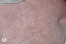
Typically presents as a morbilliform exanthem, often with perifollicular accentuation.


Initial predilection for acral sites (e.g. dorsal hands and feet, palms, soles, ears), forearms, and upper trunk.


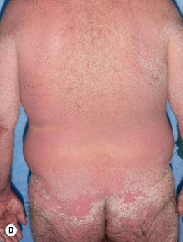
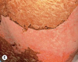
Clinical staging is based on the proportion of the cutaneous surface involved: Stage 1, <25%; Stage 2, 25–50%; and Stage 3, >50%. Stage 4 represents erythroderma with bullae/epidermal detachment resembling toxic epidermal necrolysis (Fig. 44.1).
Apr 22, 2016 | Posted by in Dermatology | Comments Off on Graft-Versus-Host Disease

Full access? Get Clinical Tree

Get Clinical Tree app for offline access