86
Giant Cell Tumor of the Tendon Sheath (Benign)
Kevin D. Plancher
History and Clinical Presentation
A 40-year-old right hand dominant woman presented with a mass on her left index finger. The patient reports that she has no symptoms related to the mass; however, occasionally she notices pain, stiffness, and numbness. The patient has had the mass for ∼2 years and does not recall any specific trauma to the area. She would like the mass removed for cosmetic reasons and because it causes her some functional problems.
Physical Examination
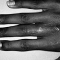
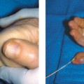
A mass is palpated on the volar ulnar aspect of the patient’s left index finger (Fig. 86–1). The mass is nontender, firm, and fixed. The skin over the mass is mobile. The mass is not transilluminating.
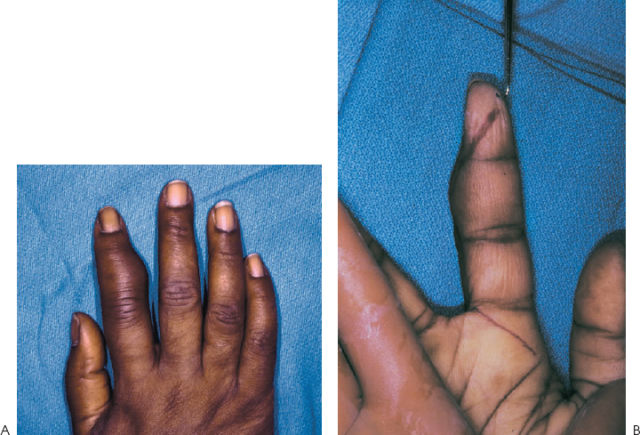
Figure 86–1. (A,B) Giant cell tumor of left index finger, ulnar aspect, with (A) dorsal and (B) volar views.
Diagnostic Studies
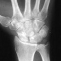
Standard radiographs show a soft tissue mass. Adjacent to the mass, cortical erosion, from the pressure effect of the mass, is seen. Bony erosion is not an uncommon finding with some giant cell tumors of the tendon sheath.
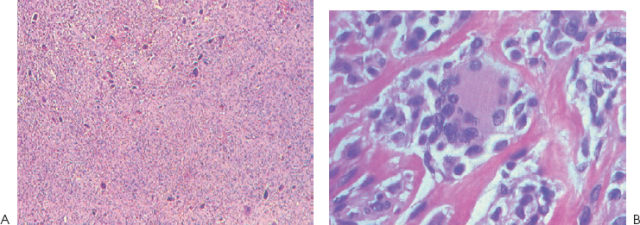
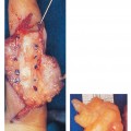
Histologic analysis reveals spindle cells, multinucleated giant cells, mononuclear cholesterol-laden histiocytes, fibroblasts, and round oval synovial cells arranged in clusters (Fig. 86–2).
Differential Diagnosis
Foreign body granuloma, necrobiotic granuloma
Related posts:
Stay updated, free articles. Join our Telegram channel

Full access? Get Clinical Tree


