10
Frontal Sinus Fractures
The frontal bone is the strongest bone of the face; a direct isolated high-energy impact is usually needed to fracture this bone. The frontal sinuses are absent in 4% of individuals, rudimentarily developed in 5%, and unilateral in 10% of individuals.
 Anatomy
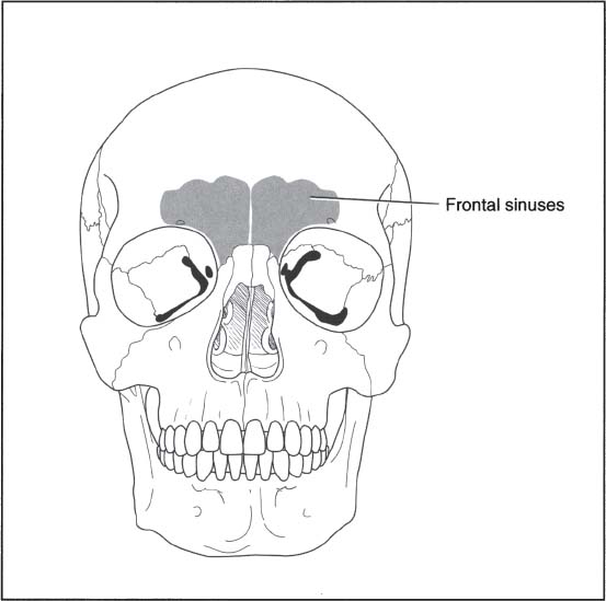
The anatomy of the frontal sinuses is comprised of (Fig. 10–1)
• Two paired irregular cavities
• Anterior wall = anterior table
• Posterior wall = posterior table
 Physical Examination
• Forehead contusion
• Forehead laceration
• Forehead or orbital hematomas
• Epistaxis
• Otorrhea or rhinorrhea from dural tears – test with halo sign on paper towel; send fluid for glucose and β transfer
• Palpable step deformity secondary to underlying fracture; may be obscured by overlying swelling in the acute setting
• Paresthesias in the supraorbital nerve distribution

Figure 10–1 The frontal sinuses.
Stay updated, free articles. Join our Telegram channel

Full access? Get Clinical Tree


