Free functional muscle transfers are an excellent treatment option in patients when significant time has passed after a nerve injury. In addition, they are the treatment of choice for reconstruction of established Volkmann’s ischemic contracture, muscle loss from trauma, or tumor resection, and in congenital muscle absence. In cases where there is both soft tissue and functional muscle loss, free functional muscle transfers can address these problems together. This article focuses on the key principles for functional reconstruction of the upper extremity with free functional muscle transfers.
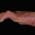
Injuries to the brachial plexus, or those to the upper extremity resulting in Volkmann’s ischemic contracture, can cause devastating functional loss. Nerve transfers for brachial plexus and peripheral nerve injuries have become the cutting-edge reconstructive options for loss of distal function in the upper extremity. Nerve transfers, direct nerve repair, or nerve grafting should be used in acute cases of plexus and/or peripheral nerve loss during the first 6 to 9 months following injury; this can allow adequate time to reinnervate distal motor targets. It is known that by 18 to 24 months, irreversible changes in muscle cells occur and limit the chance of motor recovery. Free functional muscle transfers are an excellent treatment option in patients when significant time has passed after a nerve injury. In addition, they are the treatment of choice for reconstruction of established Volkmann’s contracture, muscle loss from trauma or tumor resection, and in congenital muscle absence. These procedures are indicated only if functional deficits cannot be restored by local muscle rotation or tendon transfers. In cases where there is both a soft tissue and functional muscle loss, free functional muscle transfers can address these problems together. The focus of this article is to highlight the key principles for functional reconstruction of the upper extremity with free functional muscle transfer.
History
In 1970, Tamai and colleagues, using a canine model, were able to successfully transfer a rectus femoris muscle to the forelimb. Free functional muscle transfer (FFMT) was first done clinically by Harii and colleagues for facial reanimation using the gracilis muscle. At the same time, surgeons at Six People’s Hospital in Shanghai transferred a portion of the pectoralis major muscle to the forearm to restore finger flexion in a patient with Volkmann’s Ischemic Contracture. In Japan, Ikuta and colleagues, used free functional gracilis muscle transfers for the same functional reconstruction. In North America, credit should be given to Manktelow and Zucker for pioneering FFMTs. Since the reports in the 1970s, the literature has increased with an expanded application of FFMT demonstrating improvement in function of the upper extremity. In recent publications, larger case series have been reported that include functional outcomes.
Patient selection
Reconstructive surgeons involved in the care of patients who may benefit from FFMT should first consider less complicated options for reconstruction. These procedures would commonly include local muscle and/or tendon transfers. In severe cases of Volkmann’s ischemic contracture, often the flexor and extensor compartments are damaged to a varying degree, limiting the local reconstructive options. In these situations a FFMT is the best option. Other patients who would potentially benefit from FFMT include, those with muscle loss from direct trauma, electrical injuries, long-standing neurologic injury, congenital absence and, now with the increase in limb salvage surgery, those with functional deficits after tumor excision ( Box 1 ).
Volkmann’s ischemic contracture
Direct traumatic loss of muscle
Electrical injury involving the upper extremity
Long-standing neurologic injury
Upper extremity tumor excision
Congenital muscle absence
For all of these indications, a compliant and motivated patient is perhaps the most important component to successful functional transfers, as the rehabilitation programs can be complex and time consuming. Patients and families must have detailed explanations of the commitment, as well as, realistic expectations for outcomes. Patients should be encouraged to meet with other patients that have had the same procedure and have successfully completed rehabilitation. It is equally important to ensure the patient does not have any other underlying medical conditions that would compromise the functional transfer or put the patient at risk by performing these procedures. In the authors’ experience, the most successful outcomes with FFMT have been when the patients are younger than 45 years of age.
Principles
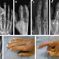
The common problem for patients who require FFMT is loss of function of one muscle, or a group of muscles. The most important reconstructed function in the upper extremity is elbow flexion. In addition, FFMTs are also used for deltoid reconstruction, elbow extension, and wrist and finger flexion and extension. With functional loss in the forearm, it is important to evaluate both the flexor and extensor muscles to determine whether an antagonistic force exists to the proposed transferred muscle. If there is no opposing muscle function, the patient may require a double transfer, or other procedures, such as wrist arthrodesis or tenodesis, to obtain this balance. The authors agree that finger extension should be reconstructed prior to finger flexion to allow for a more rapid return of function and faster rehabilitation. A prerequisite for a successful FFMT is a healthy recipient bed to allow tendon gliding. If this does not exist, the patient may require a staged procedure to prepare the arm for an FFMT, such as, local tissue rearrangement, tissue expansion, or free tissue transfer.
Success also depends on the patients having mobile joints and gliding tendons. If this mobility does not exist, the patient requires contracture release, tenolysis, and/or capsulotomies. It is essential to combine these initial procedures with aggressive physiotherapy, to achieve full passive range of motion before the planned FFMT ( Box 2 ).
Good soft-tissue coverage for the reconstruction site
Full passive range of motion of the joints the transfer will act across
Pure, undamaged donor motor nerve at the site of transfer
Reliable recipient vessels
Adequate tendon glide
Antagonistic muscle function
Motivated, compliant patient
Adequate physical therapy
Viability of the transferred muscle is achieved using meticulous microsurgical technique. Successful FFMT requires a pure motor nerve to power the transfer with an appropriate size match to the donor nerve. The nerve coaptation should be as close as possible to the transferred muscle to minimize the reinnervation distance. Good sensation of the hand is important for optimal functional results, and this often may require sensory nerve reconstruction at the same time as, or before, the motor reconstruction. To ensure successful FFMT, the resting length of the muscle must be correctly restored. These key principles are summarized in Box 3 .
Plan incisions for exposure and tendon coverage
Prepare tendon for muscle insertion with normal cascade
Select healthy vessels close to the muscle pedicle
Select healthy motor nerve
Perform a nerve repair as close to muscle as possible to minimize time of denervation
Secure fixation at origin and insertion to minimize stretching
Ensure correct resting length of the muscle
FFMT requires a great deal of preoperative planning; however, the care of these patients requires a multidisciplinary team with the ability to support the intensive and long postoperative rehabilitation.
Muscle selection
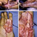
There have been many muscles studied for their suitability to be used as FFMT ( Box 4 ). Desirable characteristics include a reliable neurovascular pedicle, as well as, having minimal or no donor site deficit. The muscle should have adequate strength and have a range of excursion that will meet the needs of the site to which it is being transferred ( Box 5 ). A good example meeting these criteria is the gracilis muscle, which is the most commonly transferred muscle to the upper extremity because of its functional and anatomic fit. The muscle can be used for deltoid reconstruction, elbow flexion, as well as finger flexion and extension. A review of 71 free gracilis functional transfers, supports the consensus that this muscle is useful for reconstruction of both finger flexion and extension. This muscle is also commonly used for reconstruction of elbow flexion. Kay and colleagues reviewed 33 patients that underwent free functional gracilis transfers for elbow flexion over a 14-year period. In their series, 70% of the patients obtained a successful result with elbow flexion M3 or greater. Using intercostal nerves as donor nerves in their series appeared to have better outcomes than using other nerves, such as ulnar fascicles or the spinal accessory nerve.
Gracilis
Latissimus dorsi
Rectus femoris
Tensor fascia lata
Gastrocnemius
Serratus anterior
Gracilis with adductor longus
Desirable neurovascular anatomy
Adequate strength
Suitable range of excursion
Suitable gross anatomy to fit defect
Adequate fascia or tendon to allow secure attachment
Minimal donor deficit and limited cosmetic donor defect
There have been many techniques described for reconstruction of elbow flexion. When no local option, such as latissimus dorsi and pectoralis major pedicled transfers are available, or the use of a proximal advancement of the flexor-pronator origin (Steindler procedure), FFMT remains the only choice. Options for restoring elbow flexion with FFMT include gracilis, latissimus dorsi, or rectus femoris muscles. In the authors’ practice the gracilis muscle is most commonly used, followed by the latissimus dorsi. In 2010, Muramatsu and colleagues published their case series, which included 7 patients requiring reconstruction after oncologic sarcoma resection in the upper extremity using free latissimus dorsi, with good results. Kay and colleagues highlight the importance of a learning curve for FFMT.
Muscle Physiology
An understanding of the length-tension curves for muscle contraction is required when planning functional muscle transfers. The contraction of a muscle is a dynamic process involving coordinated contraction of thousands of muscle fibers consisting of overlapping actin and myosin filaments. The force of contraction generated by a muscle is directly proportional to the amount of overlap of these thick and thin fibers, in a calcium-dependent process known as the sliding filament mechanism of contracture. This process is influenced by the length the muscle is stretched before it contracts. With the muscle stretched beyond its normal limits, there is the least amount of overlap between the actin and myosin fibers, and therefore the muscle contraction is weak. As the muscle shortens and the amount of overlap is increased between the fibers, the strength of muscle contraction increases to a maximum point found at the resting length in its most elongated position. This point is the peak of the length-tension curve for the muscle after which, if the muscle continues to shorten, there is less optimization of the overlap of myosin fibers relative to actin, resulting in less force of muscle contraction. The goal of tensioning a functional muscle transfer is to allow the muscle to act at its peak length-tension point for contraction at its resting length.
It must be noted that the connective tissue framework surrounding the muscle fibers contributes to the contraction force. The connective tissue provides a recoil tension when the muscle is in full extension, adding to the contractile force, and limiting the maximal extension of the muscle. Muscle contraction relates to the number of muscle fibers that are activated by a neural stimulus, and contraction strength is increased as more fibers are recruited to contract. Maximal innervation of the muscle is therefore important for functional transfers to ensure that the maximum strength is obtained from the muscle.
Vascular considerations
It is of paramount importance to use meticulous technique for the vascular anastomoses for FFMT, and the reconstructive surgeon must carefully plan the location of these to ensure there is no tension on the pedicle with muscle excursion. Standard options for vascular anastomosis include end-to-end suturing to existing, healthy named vessels. If there is a large mismatch between the donor and recipient vessels, end-to-side anastomosis should be performed.
For successful FFMT, the recipient vessels ideally should be of equivalent caliber to the donor vessels. In addition, the anastomosis should be in healthy tissue away from the site of injury. The vena comitantes in the upper extremity, especially in cases of previous trauma and ischemic injuries, can often be damaged. Venous outflow problems are more common than arterial inflow problems for complications with FFMT; therefore, the vena comitantes should always be flushed with heparinized saline to ensure they are draining well. If there is any resistance in these veins, other deep veins or superficial veins should be used. It is important to look for and protect superficial arm veins during the initial incisions and flap elevation in the upper arm and forearm for this reason. When using superficial veins there is often a mismatch in size between the donor and recipient vein, making them more difficult to use.
The most common vascular pedicle used at the shoulder for deltoid reconstruction, or elbow flexion, is the thoracoacromial artery, with either the associated vena comitantes or a branch of the cephalic vein. This is due to the excellent size match between the donor and recipient vessels and its anatomic location. Alternatively, the thoracodorsal artery and vena comitantes can be used.
For finger flexion and extension reconstruction, if the ulnar artery is present the authors’ preference is to use the radial artery and vena comitantes. If it is not present, the anterior interosseous artery is a viable option, or an end-to-side anastomosis to the radial artery can also be performed.
Nerve considerations
A preoperative physical examination should always be performed to identify which nerves are functioning in the upper extremity. For successful FFMT, the donor nerve must be a pure motor nerve, with no surrounding scar tissue, and ideally with a synergistic function to the muscle the transfer is replacing. If there is any question on the viability of the donor nerve, an intraoperative biopsy should be performed to confirm its health. This action is undertaken before the functional muscle is harvested.
For deltoid or elbow flexion reconstruction, preference is to use either the musculocutaneous or axillary nerves if they are present. Alternatively, the spinal accessory nerve or 3 intercostal motor nerves can be used, usually the third, fourth, and fifth branches. Chuang and colleagues found that if the original musculocutaneous nerve is present, the results are better than intercostal reinnervation. It is important to perform direct repair without intervening nerve grafts to maximize the functional outcome. For triceps reconstruction and elbow extension, a branch of the radial nerve to the long head of the triceps is preferred. If this is not present, motor fascicles from either the median or ulnar nerve can be used.
The anterior interosseous nerve is the best choice for reconstruction of finger flexion. If this is not a viable option, fascicles of the median nerve to the flexor digitorum superficialis (FDS), or a fascicle of the ulnar nerve to the flexor carpi ulnaris muscle, can be used if available. For finger extension reconstruction, the posterior interosseous nerve is ideal. If this is not possible, again, branches of the median nerve to the FDS can be used.
In patients with complete brachial plexus palsy, the only motor nerve options for reconstruction on the side of injury are the spinal accessory nerve, intercostal nerves, and phrenic nerve. In the authors practice, only the spinal accessory and intercostal nerves are used. The authors agree with other investigators that it is important to keep in mind that when there is preceding chest trauma, these nerves should be used with caution. In a case series of elbow reconstruction presented by Kay and colleagues, a better motor outcome was achieved when the intercostal nerves were used as donor nerves as opposed to ulnar fascicles.
Other investigators have reported successfully using the contralateral C7 and the contralateral medial pectoral nerve with nerve grafting for reconstruction. In a modest series of a dozen cases using these two procedures, the authors were unable to obtain useful functional outcomes, and because of this, in their practice, these procedures are no longer used. This result is supported by Terzis and Kostopoulos, who in their recent review of 74 functional muscle transfers found that for finger flexion, intercostals, upper ipsilateral plexus, and distal accessory gave better results than the contralateral C7.
Preoperative planning
The preoperative planning for these patients starts with a detailed history outlining the mechanism of injury, previous investigations, interventions, and the remaining functional impairment of the patient. A detailed physical examination should include the vascular system, as well as, evaluation of the nerves, muscles, joints, and soft tissues of the upper extremity.
Formal nerve conduction studies are often not indicated or helpful. Electromyography (EMG) can be useful to assess muscle function. In the distal forearm EMG evaluation of the pronator quadratus can give information about the status of the anterior interosseous nerve function.
Magnetic resonance imaging (MRI) is required to assess viability of the affected muscles in the upper extremity. With the newer higher-powered MRI studies (3 Tesla and above), information can be obtained about the conditions of the nerves, and this can help for preoperative planning and decisions on the need for nerve grafting to restore sensation.
Angiography is recommended to provide valuable information about the arterial inflow for the extremity, especially after significant traumatic injuries. It is useful to assess interosseous vessels in these studies, as these can serve as key recipient vessels for functional transfers. Often an intact anterior interosseous artery will correlate with an uninjured anterior interosseous nerve.
Muscle selection is based on the functional requirements at the recipient site; these include the muscle length and expected excursion. Ideally the transferred muscle will span from a selected origin to insertion, optimizing its resting length. The muscle should also have adequate fascia and tendon to facilitate a secure attachment to the origin and insertion. The vascular and nerve anatomy should be considered to ensure the orientation to the recipient vessels and nerve is optimized. Finally, the functional and cosmetic deficit of the donor site should be minimized.
Adequate soft-tissue coverage is essential for a successful transfer, and may require staging the reconstruction with a first-stage local, regional, or free tissue transfer. It is always preferable to use a myocutaneous flap for the functional transfer. The benefit of this is it provides a reliable skin paddle to monitor the muscle postoperatively, and, in addition, provides a healthy gliding surface and coverage over the muscle. Myocutaneous flaps can, however, be bulky, and result in a less desirable cosmetic result. Skin grafts over the muscle belly proximal to the musculocutaneous junction can be used initially and heal well cosmetically; however, the authors recommend these to be replaced secondarily and covered with a skin flap to improve gliding. Incisions should always be planned for access and exposure of the donor vessels and nerves to facilitate the microsurgery, as well as, to ensure adequate coverage for the musculotendinous junctions.
Related posts:

Stay updated, free articles. Join our Telegram channel

Full access? Get Clinical Tree


