Fig. 5.1
Supraorbital foramen. Reproduction of a lithograph plate from Gray’s anatomy (Henry Gray, Anatomy: Descriptive and Surgical)
Fig. 5.2
Supraorbital nerve and vessels and supratrochlear nerve are shown during the dissection (source: wikipedia, www.anatomyumftm.com)
Fig. 5.3
Supratrochlear (yellow arrow) and supraorbital (green arrow) nerves and vessels have been schematically drown on the patient. Reproduction of a lithograph plate from Gray’s anatomy (Henry Gray, Anatomy: Descriptive and Surgical)
Knowledge of mimic muscle of glabellar and frontal area together with their function is of capital importance to correct esthetic issues in this region.
The frontalis muscle (Fig. 5.4) is thin, of a quadrilateral form, and intimately adherent to the superficial fascia. It has no bony attachments. Its medial fibers are continuous with those of the procerus; its immediate fibers blend with the corrugator and orbicularis oculi, thus attached to the skin of the eyebrows; and its lateral fibers are also blended with the latter muscle over the zygomatic process of the frontal bone.
Fig. 5.4
Frontalis muscle. Reproduction of a lithograph plate from Gray’s anatomy (Henry Gray, Anatomy: Descriptive and Surgical)
In the eyebrows, its primary function is to lift them (Fig. 5.5) (thus opposing the orbital portion of the orbicularis), especially when looking up.
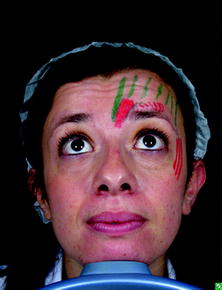
Fig. 5.5
Frontalis muscle (green) in function
From these attachments, the fibers are directed upward and join the galea aponeurotica below the coronal suture. The medial margins of the frontalis are joined together for some distance above the root of the nose.
The procerus (Fig. 5.6) is a small pyramidal slip of muscle which arises by tendinous fibers from the fascia covering the lower part of the nasal bone and upper part of the lateral nasal cartilage.
Fig. 5.6
Procerus muscle
It is inserted into the skin in the lower part of the forehead between the two eyebrows on either side of the midline, its fibers merging with those of the frontalis.
It helps to pull that part of the skin between the eyebrows downwards, which assists in flaring the nostrils.
It can also contribute to an expression of anger (Fig. 5.8).
The corrugator supercilii (Fig. 5.7) is a small, narrow, pyramidal muscle, placed at the medial end of the eyebrow, beneath the frontalis and just above orbicularis oculi.
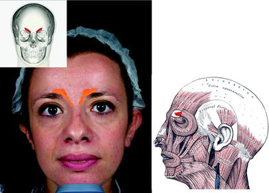
Fig. 5.7
Corrugator supercilii
It arises from the medial end of the superciliary arch, and its fibers pass upward and laterally, between the palpebral and orbital portions of the orbicularis oculi, and are inserted into the deep surface of the skin, above the middle of the orbital arch. The corrugator draws the eyebrow downward and medially, producing the vertical wrinkles of the forehead (Fig. 5.8). It is the “frowning” muscle and may be regarded as the principal muscle in the expression of suffering.
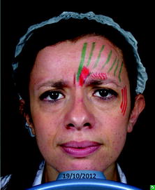
Fig. 5.8
Procerus and corrugator supercilii in action; they are usually activated in the same time
The orbicularis oculi is a muscle in the face that closes the eyelids (Fig. 5.9, 5.10). It arises from the nasal part of the frontal bone, from the frontal process of the maxilla in front of the lacrimal groove, and from the anterior surface and borders of a short fibrous band, the medial palpebral ligament.
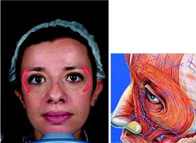
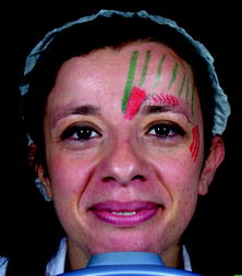
Fig. 5.9
Orbicular oculis
Fig. 5.10
Orbicular oculis in function
From this origin, the fibers are directed lateralward, forming a broad and thin layer, which occupies the eyelids or palpebrae, surrounds the circumference of the orbit, and spreads over the temple, and downward on the cheek.
The lateral part of orbicularis functionally responsible of the crow’s-feet lies above the lateral orbital rim and superficially to the temporalis muscle.
5.2 Correction of Forehead and Glabella Wrinkles and Crow’s-feet with Botulinum Toxin
5.2.1 Pitfalls
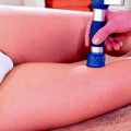
Technical skill: The goal is to inject orbicularis muscle to correct crow’s-feet. For this reason, injection should be administered immediately under the epidermis because the orbicularis muscles addressed are extremely superficial at this site. Deeper infiltration will lead to reach the layer of temporalis muscles that lies underneath the orbicolaris muscle, with consequent waste of product and insufficient esthetic result (Fig. 5.11a, b).
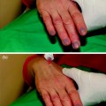
Fig. 5.11
a, b It is always useful to determine the anatomic boundaries individuating the muscles before injecting: the patient may be asked to contract frontalis muscle, procerus, corrugator, and orbicularis oculi to tailor the treatment to the anatomic features. Since the patient is young, a small quantity of toxin is injected. Six points of injection are administered on both side plus one central point as shown by the pictures. In the central point, 0.05 cc of bocouture is injected, while in all the other points just 0.5 of the solution contained in the syringe is delivered: in terms of units, 4 units in the central point and 2 units in all the other points. The other six points are: an ideal point corresponding vertically to the internal canthus placed 0.5 cm above the superior orbital rim; an ideal point corresponding vertically to the centre of the pupil placed 1 cm above the superior orbital rim; the superomedial part of frontalis muscle; the superolateral part of frontalis muscle. The orbicularis oculi is injected into three points keeping a distance of 1 cm from the lateral orbital rim. The described safepoints are indicated as yellow circles on patient’s face
Safe points: While injecting, a good manner is to touch with the hand that does not handle the syringe the bone rim of the orbit, ensuring this way that botulinum is delivered in the safe points: in particular, one should never inject to close to the eyebrows (0.5 cm distance at least, 1 cm in the medium part of the eyebrows) to avoid the possibility of diffusion to levator muscle and subsequent upper eyelid ptosis.
5.2.2 Indications
Official indication for aesthetic use of botulin toxin is the temporary improvement in the appearance of moderate to severe glabellar lines (vertical lines between the eyebrows) seen at frown, in adult patients under 65 years, when the severity of these lines has an important psychological impact on the patient.
Other indications are off label, and these are the temporary improvement of crown feet and of forehead lines at rest and in motion.
5.2.3 Contraindications
Contraindications to treatment with BoNT/A are as follows:
diseases of the neuromuscular junction (myasthenia gravis, Lambert–Eaton syndrome);
allergy to human albumin and sodium chloride;
skin lesions of treated areas;
infection of the planned site for the injections;
recent surgical treatments;
pregnancy and lactation;
recent use of aminoglycoside antibiotics or other agents that interfere with neuromuscular transmission (spectinomycin, tubocurarine-type muscle relaxants).
5.2.4 Case 1: Treatment with Bocouture in a Young Simmetric Patient
Materials (Fig. 5.12 ).
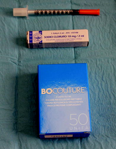
Fig. 5.12


Related posts:


Stay updated, free articles. Join our Telegram channel
Full access? Get Clinical Tree
