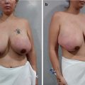
Systemic manifestations
Local manifestations
Myalgia, myositis, muscle weakness
Inflammatory nodules
Arthralgias and/or arthritis
Cutaneous hyperpigmentation, edema and angioedema
Pyrexia, dry mouth, chronic fatigue
Cutaneous induration, pseudo abscesses
Sleep disorders, cognitive disorders
Axillary lymphadenopathy
Palmar erythema, livedo reticularis
Panniculitis, morphea, and sarcoidosis
Neurological manifestations
Necrosis, ulcers
Pulmonary manifestations, pulmonary hypertension, dyspnea
Systemic lupus erythematosus (SLE)
Vasculitis
Sjogren’s syndrome
Scleroderma
Eosinophilic fasciitis
Autoimmune syndrome induced by adjuvants (ASIA)
Still’s disease
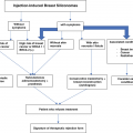
It should be noted that eliminating the triggering agent improves symptoms. Recently, Shoenfeld and Agmon-Levin coined the term adjuvant-induced autoimmune/autoinflammatory syndrome (ASIA) to define a set of conditions that result from a body’s innate immune response to adjuvants (aluminum, silicone, bacterial antigens, etc.) (see Chaps. 6 and 8) [6].
Silicone has an adjuvant effect and can act as a pathogen-associated molecular pattern, resulting in the activation of pattern-recognition receptors and the release of proinflammatory cytokines, like TNF-α, IL-1β, IL-6, IFN-γ, and IFN-α [7–9]. Of these, the last is responsible for inducing the maturation of monocytes in dendritic cells, increasing the release of co-stimulatory molecules, and triggering the expression of the major histocompatibility complex which, aided by IL-6, favors the secretion of antibodies [10, 11]. In addition to activating the immune system, silicone can cause systemic effects via the degradation of its fragments. In patients with severe immune reactions, there is an increased concentration of immunoglobulin G in surrounding tissues and high levels of anti-silicone antibodies.
Diagnostic Imaging
Diagnostic utility of imaging studies
Mammography | Breast ultrasound | MRI |
|---|---|---|
Requirements: Perform bilateral examinations, in craniocaudal and mediolateral oblique positions. Include the axillae | Requirements: The examination should extend beyond the limits of the breast and include the chest wall and axilla | Requirements: The examination should be performed with the patient in a prone position. Administer gadolinium IV. Use high magnetic field equipment (1,5 or 3 T) and specific coils. This allows one to explore both breasts with extension into the chest wall and axilla |
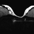
Strengths: Greater sensitivity to detect liquid silicone in breast parenchyma (in all mammary planes, from subdermal to posterior). It also shows infiltrates in the pectoral muscle and axillae. It is possible to observe compromise in the lymphatic tissue (expressed as dense, linear and parallel lines, shaped like multi-lobed lobed nodules) | Strengths: The ultrasound image usually has the appearance of a “snowfall” (intense and homogenously hyperechoic nodules, with a delimited and rounded anterior contour), which generates a shadow that obscures the posterior border [12]. Some silicone collections initially present as complex cysts. It is also possible to observe isoechogenic solid nodules and fibrosis | Strengths: This technique offers morphological information and data related to breast parenchymal physiology (perfusion and enhancement kinetics). It does not use ionizing radiation. Injected silicone may manifest as nodular images or as silicone-signaling images that infiltrate the fibroglandular tissue and may migrate to the adjacent axilla and other soft tissues [12]. Additional benefits: Determines the location of silicone and its eventual migration. It allows for visualization of pathologies that cannot be observed by conventional studies. It has a very high negative predictive value for invasive carcinoma, in the absence of enhancement [12] |
Limitations: 1. The high density of siliconomas and diffuse infiltration of breast tissue make it difficult to interpret suspicious images (microcalcifications or non-visible nodules), which prevents the diagnosis of small cancerous lesions 2. It is impossible to evaluate the full extent of the siliconoma, since only that sector of the body included in the field of study can be observed, leaving out the intermammary groove, abdomen, and thoracic wall [12] | Limitations: Ultrasound is not useful to detect lesions suspected of being neoplastic | Limitations: It has high sensitivity to detect lesions, but only moderate specificity for detecting breast cancer [12] |
Related posts:

Stay updated, free articles. Join our Telegram channel

Full access? Get Clinical Tree


