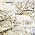
Anatomy of the lateral wall of the nose (with opened middle turbinate)
12.2.3 Operative Technique
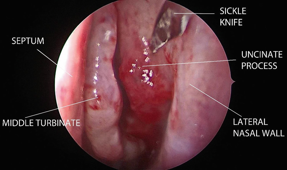
By using a 0 degree Hopkins telescope, the anterior attachment of the uncinate process is incised with a sickle knife and medialised to reveal the infundibulum. The upper and lower attachments of the uncinate process are cut with a straight Blakesley forceps, and it is removed with a twisting motion (Fig. 12.2).
Fig. 12.2
The first step in FESS is the uncinectomy
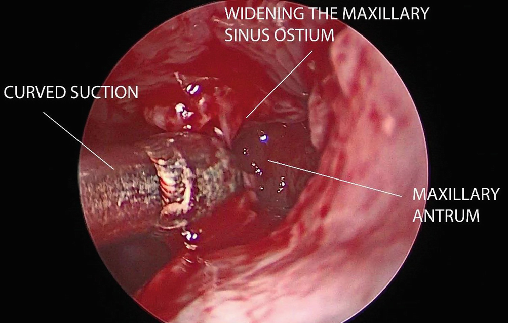
Maxillary ostium is usually visible after uncinectomy, or else it can be identified with a blunt probe, blunt curette or curved sucker or by visualising the presence of mucus. The maxillary ostium is widened posteriorly with a straight Blakesley forceps and anteriorly with a backbiting forceps (Fig. 12.3).
Fig. 12.3
Maxillary antrostomy is done by using a combination of microinstruments and microdebrider
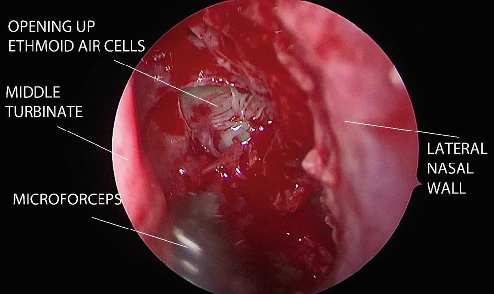
The anterior ethmoid air cells are opened up with a straight Blakesley forceps and removed in piecemeal fashion. Posterior ethmoidectomy is done by penetrating the basal lamella with ball probe at its medial and inferior part. Posterior ethmoid cells are then removed by either blunt curette or straight Blakesley forceps (Fig. 12.4).
Fig. 12.4
Ethmoidectomy is done whereby the anterior and posterior ethmoid air cells are opened up
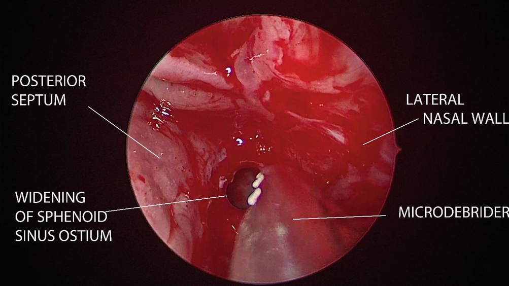
The sphenoid sinus is entered as inferiorly and medially as possible from the posterior ethmoid cell if a transethmoid sphenoidotomy is being done. The sphenoid ostium can also be identified by blunt probing 1.5 cm from the posterior choanae or by excising a small inferior part of superior turbinate when a transnasal sphenoidotomy is being done. When clearing the sphenoid sinuses of disease, the carotid artery and optic nerve located on both of its lateral wall must be kept in mind (Fig. 12.5).
Fig. 12.5
Sphenoidotomy is done by using a microdebrider
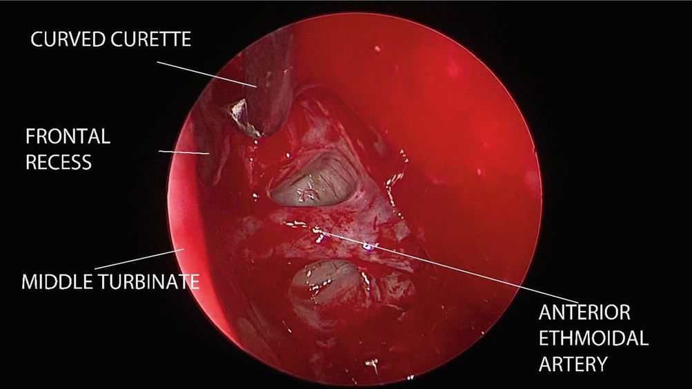
The frontal recess is opened up and cleared of disease by using a combination of a 30 or 45 degree telescope with either microinstruments or microdebrider (Fig. 12.6).
Fig. 12.6
Frontal recess is opened up by using blunt curved curette. The anterior ethmoidal artery is located at its posteroinferior border
Patients with rhinogenic headache can benefit from FESS and surgical intervention ought to be individualised based on patient’s endoscopic and imaging findings of the location of anatomical variations.
12.3 Rhinogenic Headache
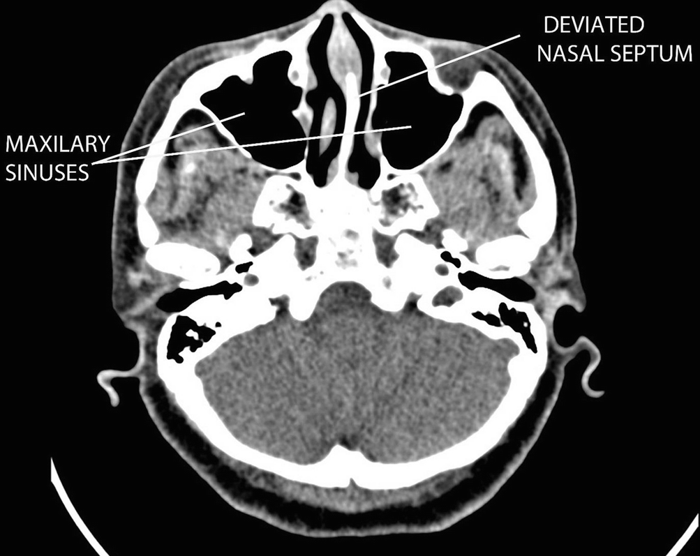
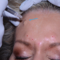
Rhinogenic headache is a headache or facial pain syndrome due to mucosal contact points in the nasal or sinus cavities in the absence of inflammatory sinonasal condition, purulent discharge, sinonasal polyps and mass or hyperplastic mucosa [2]. Neuropeptides and substance P are involved in the mediation of facial pain due to the contact between the mucosal surfaces [3]. The release of those mediators is responsible for the migraine-like headache symptoms. The treatment for rhinogenic headache is to remove or to release the contact points of the mucosal area in the nose (Fig. 12.7). This can be done via an endoscopic septoplasty and an endoscopic inferior turbinate reduction surgery (endoscopic turbinoplasty).
Fig. 12.7
CT PNS showing mucosal contact points in the nose
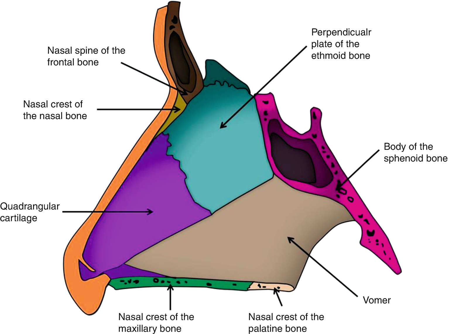
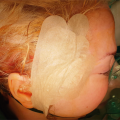
12.3.1 Anatomy of the Nasal Septum
Nasal septum separates the two nasal cavities. It is formed by a cartilaginous part anteriorly, a bony part inferoposteriorly and a small anterior membranous part, the columella. The bony part is composed of perpendicular plate of ethmoid, vomer, nasal spine of the frontal bone, rostrum of the sphenoid, nasal crest of the nasal bone, nasal crest of the palatine bone and nasal crest of the maxillary bone (Fig. 12.8). The quadrangular cartilage forms the cartilaginous part [4]. Nasal septum is the central support structure for the nose. If the septum is significantly deformed, the septum may cause dysfunction and cosmetic deformity which potentially have an impact on numerous significant functions of the nasal cavity.
Only gold members can continue reading. Log In or Register to continue
 Approach for Auriculotemporal Nerve Decompression, Amirlak Modification
 Anatomy of Craniofacial Nerves Regarding Migraine Surgery
 Regional, Targeted (ART) Botulinum Toxin Injection for Migraine and Chronic Headaches
 Anatomy of the Vascularization and Innervation of the Human Scalp
 of Temporal Trigger Sites
 of Occipital Trigger Sites


