In addition to reflectance confocal microscopy, multiwave confocal microscopes with different laser wavelengths in combination with exogenous fluorophores allow fluorescence mode confocal microscopy in vivo and ex vivo. Fluorescence mode confocal microscopy improves the contrast between the epithelium and the surrounding soft tissue and allows the depiction of certain structures, like epithelial tumors, nerves, and glands.
Key points
- •
Fluorescence confocal microscopy (FCM) improves the contrast between the epithelium and the surrounding soft tissue and allows the depiction of certain structures, like epithelial tumors, nerves, and glands.
- •
Main indications include instant margin control in basal cell carcinoma (BCC) on the excised tissue and studies on barrier function and skin protection in vivo and ex vivo.
- •
Further optimizations on FCM parameters and fluorophores are needed to improve FCM, offering in addition to the reflection mode, valuable image information for the selective imaging of certain target structures in the human skin in vivo and ex vivo.
Introduction
In reflectance confocal microscopy contrasts are achieved due to different refractive indices of cell structures and organelles. Keratin and melanin serve as endogenous chromophores in the reflection mode due to a higher refractive index than water.
With the development of so-called multiwave devices, additional lasers with wavelengths of 445/488 nm, 658 nm, and 785 nm were added to an 830-nm laser. By using exogenous fluorescent dyes and different filters, they offer a fluorescence mode in addition to the reflectance mode. Corresponding to the different laser wavelengths, each fluorophore has a certain excitation and emission maximum. Because the emitted fluorescence spectrum has longer wavelengths with respect to the absorption spectrum, it is, therefore, usually red-shifted. The reflection light is much brighter than the fluorescence light, so filters are needed to keep out the reflection light and to gather only the fluorescent light. Fluorescence microscopy can be performed with a laser light source with correct wavelength and laser power, a filter system allowing for display of reflection, fluorescence, or both, and fluorophores matching these parameters.
The range of possible indications for FCM is growing rapidly. FCM is used in vivo for studies on skin barrier function, nanocarriers, and penetration. For ex vivo FCM, the main indication is instant tumor margin control of BCCs during Mohs micrographic surgery by increasing the contrast between epithelial tumor and stroma.
This article offers information on the staining modalities in FCM. Suitable fluorophores for the available laser wavelengths with corresponding excitation and emission maxima, their applicability in vivo and ex vivo in different concentrations (especially in terms of toxicity), and how to overcome problems, such as photobleaching, quenching, and pH sensitivity (specific and different for each particular dye), are reviewed.
Content
Devices
The multilaser confocal microscopes suitable for FCM are the VivaScope 1500 Multilaser for in vivo and the VivaScope 2500 Multilaser for examination of freshly excised tissue (MAVIG, Munich, Germany). Both devices offer cellular resolution. In vivo, the penetration depth is limited to approximately 250 μm to 300 μm. For the ex vivo examination, there is no limitation to the depth as the tissue is placed on the cutting edge. Depending on the wavelength of the different lasers and dependent on the features of the devices (filters and so forth), reflection, fluorescence, or both are possible. Detailed technical data of both devices are as follows (new parameters of the latest device generation in parentheses).
VivaScope 1500 Multilaser for in vivo use
- •
Reflection mode wavelengths 445 (488) nm, 658 nm, and 830 (785) nm
- •
Fluorescence mode wavelengths 445 (488) nm, 658 nm, and 830 (785) nm
- •
Filter position only reflection, only fluorescence, both reflection and fluorescence
- •
Field of view 0.5 mm × 0.5 mm up to 8 mm × 8 mm
- •
Optical resolution horizontal less than 1.25 μm, vertical less than 5 μm
VivaScope 2500 Multilaser for ex vivo use
- •
Reflection mode wavelength 830 nm
- •
Fluorescence mode wavelengths 445 (488) nm and 658 nm
- •
Filter position fixed: 830-nm only reflection, 445 (488) nm, and 658-nm only fluorescence
- •
Field of view 0.75 mm × 0.75 mm (0.63 mm × 0.63 mm) up to 12 mm × 12 mm (20 mm × 20 mm)
- •
Optical resolution horizontal less than 1.25 μm, vertical less than 5 μm
Fluorophores
For the optimal dye, certain properties have to be fulfilled: excitation and emission wavelength have to correspond with the given lasers and filters. Tissue toxicity and patient safety have to be considered. Best concentrations for in vivo and ex vivo use and adequate staining procedures have to be established. Also, further biological and chemical properties, such as stability, specificity of binding, and membrane penetration, have to be taken into account. Last, but not least, important properties, such as availability and prize, should not result in major restrictions.
Fluorescein, acridine orange, methylene blue, patent blue, Nile blue, and indocyanine green (ICG) were identified as suitable candidate fluorophores according to their excitation maxima (supplier in parentheses):
445 (488) nm
- •
Fluorescein (Alcon Pharma, Freiburg, Germany), approved for angiography of the eye
- •
Acridine orange (Sigma-Aldrich Chemie, Munich, Germany), approved for ex vivo use
- •
658 nm
- •
Methylene blue (Sigma-Aldrich Chemie), approved as a drug
- •
Patent blue (Guerbet, Sulzbach, Germany), approved for lymphatic mapping
- •
Nile blue (Sigma-Aldrich Chemie), approved for ex vivo use
- •
758 nm
- •
ICG (Pulsion Medical Systems, Feldkirchen, Germany), approved for angiography of the eye
- •
Depending on their tissue toxicity, the fluorophore can be used in vivo (topically or intracutaneously) and/or ex vivo in different concentrations.
In Vivo Fluorescence Confocal Microscopy
The issue of patient safety is the most important one for in vivo examinations. ICG is approved for in vivo use but is generally also toxic to tissue and can cause pain and necrosis; the authors have used it intradermally in low concentrations. Without restrictions in terms of toxicity and therefore appropriate for in vivo use are fluorescein, patent blue and methylene blue, although the latter bears the risk of permanent tattooing.
Due to the barrier function of the skin, different results can be expected, if the fluorophores are applied topically or intracutaneously in vivo.
Topical application
To mark the superficial layers of the stratum corneum, topical application of the dye is sufficient. The dye is dropped on the skin surface and wiped off with a paper cloth.
Fluorescein, methylene blue, patent blue V, and ICG are suitable for topical application.
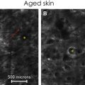
Intradermal injection
To look at the lower epidermal layers and the upper dermis and to omit the barrier function of the stratum corneum, fluorescein or ICG can be injected intradermally after disinfection with a standard alcoholic disinfectant. ICG has to be used with caution because it is not approved for intradermal use, and tissue necrosis and pain at the injection site are possible. In the authors’ studies, discoloration of the injection site for more than 24 hours and slight pain during injection were the only noticed side effects ( Fig. 1 ).