(1)
Hôpital Universitaire de Strasbourg, Strasbourg, France
Abstract
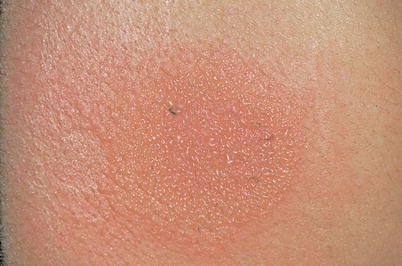
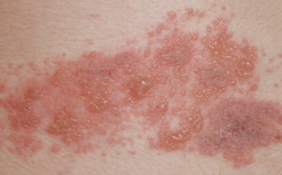
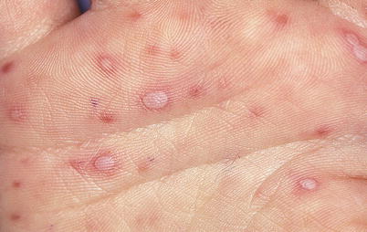
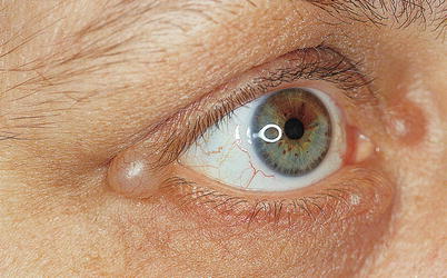
Vesicles are lesions of less than 5 mm that contain (clear) fluid (Fig. 5.1). In order to distinguish vesicles from small papules of solid content, it is sometimes necessary to puncture the top of a vesicle by means of a vaccinostyle (a pointed lancet) or a small needle, to make sure it is fluid-filled. Vesicles are sometimes conspicuous and produce a translucent lesion, which can be round (hemispherical), conical (acuminate), or depressed (umbilicate) (see Fig. 2.11). However, they are often fragile and temporary and can burst and produce weeping, erosions, and crusting, borders of crusts being rounded, fragmented, or polycyclic. Microscopic vesicles may produce clinical aspects appearing as erythematosquamous or papular. Vesicles evolving towards weeping usually indicate a spongiosis from the histopathological point of view; they are typical of eczema (“dermatitis”). Conversely, vesicles evolving towards umbilication and/or necrosis without weeping often reflect a viral cytopathogenic effect, as in infections caused by herpesvirus or zoster/varicella virus (Fig. 5.2). Oblong vesicles found on extremities are characteristic of hand-foot-and-mouth syndrome (Fig. 5.3). A vesicle should not be mistaken with a hidrocystoma (Fig. 5.4), a translucent sudoral cyst, or pseudovesicles of lymphedema (Fig. 5.5).
5.1 Vesicle
Vesicles are lesions of less than 5 mm that contain (clear) fluid (Fig. 5.1). In order to distinguish vesicles from small papules of solid content, it is sometimes necessary to puncture the top of a vesicle by means of a vaccinostyle (a pointed lancet) or a small needle, to make sure it is fluid-filled. Vesicles are sometimes conspicuous and produce a translucent lesion, which can be round (hemispherical), conical (acuminate), or depressed (umbilicate) (see Fig. 2.11). However, they are often fragile and temporary and can burst and produce weeping, erosions, and crusting, borders of crusts being rounded, fragmented, or polycyclic. Microscopic vesicles may produce clinical aspects appearing as erythematosquamous or papular. Vesicles evolving towards weeping usually indicate a spongiosis from the histopathological point of view; they are typical of eczema (“dermatitis”). Conversely, vesicles evolving towards umbilication and/or necrosis without weeping often reflect a viral cytopathogenic effect, as in infections caused by herpesvirus or zoster/varicella virus (Fig. 5.2). Oblong vesicles found on extremities are characteristic of hand-foot-and-mouth syndrome (Fig. 5.3). A vesicle should not be mistaken with a hidrocystoma (Fig. 5.4), a translucent sudoral cyst, or pseudovesicles of lymphedema (Fig. 5.5).


Fig. 5.1
Vesicles. Acute dermatitis (eczema). Multiple confluent and translucent vesicles on an erythematous background, as observed in acute allergic contact dermatitis. A vesicle is a fluid-filled lesion of less than 5 mm. It is one of the primary lesions of dermatitis. The usual course of dermatitis includes erythema, vesicles, weeping erosion, crusting, and recovery. Intense pruritus is present at all stages. The absence of pruritus requires that the diagnosis be revised. The vesicle observed in dermatitis is the prototype example of vesicles that develop weeping, which reflects the pathogenic mechanism of spongiosis. Spongiosis indicates intercellular epidermal edema. Viral vesicles produced by other mechanisms (necrosis) do not evolve towards weeping

Fig. 5.2
Vesicles. Zoster. Grouped vesicles on an erythematous background. Several elements are characteristic of the viral nature of the vesicles on one side and of its cause, zoster, on the other. The gray color and the umbilication are typical of the viral nature. Clustering is typical of herpetic infections. The metameric arrangement immediately indicates zoster (shingles). Unlike vesicles of dermatitis (eczema), these lesions do not evolve towards weeping but may evolve into a painful cutaneous necrosis

Fig. 5.3
Oblong vesicles. Hand-foot-and-mouth disease. The gray color is characteristic of a viral lesion. Note the oblong shape and the erythematous border of these lesions. In this disorder, the lesions are located on the palms and soles. The disorder itself is caused by a Coxsackie virus infection (benign form) or an echovirus (more serious form due to neuromeningeal and cardiopulmonary risks). Buccal vesicular erosive involvement may also exist

Fig. 5.4


Differential diagnosis of a vesicle. Telangiectatic translucent papule. Hidrocystoma. This is a thin-wall sudoriparous cyst located in the superficial dermis. This cyst may resemble a vesicle and can be emptied through puncturing. Its fluid is however more viscous. It is almost always located around the eyes
Stay updated, free articles. Join our Telegram channel

Full access? Get Clinical Tree


