(1)
Hôpital Universitaire de Strasbourg, Strasbourg, France
3.1 Macule
3.2 Erythema
3.3 Cyanosis
3.4 Telangiectases
3.5 Poikiloderma
3.6 Purpura
3.7 Atrophic Macule
3.8.1 Erythema and Angioma
3.8.2 Purpura
3.8.3 Telangiectasia
3.8.4 Cyanosis
3.8.5 Livedo
3.8.6 Color of Macules
Abstract
When a lesion is not palpable, it becomes apparent because of localized skin discoloration. These lesions are usually called macules when they measure less than 2 cm and patches when they are larger. This distinction between macules and patches, based on diameter, has no practical interest and therefore the term macule will be used to refer to localized changes in skin color, regardless of size. Macules are classified according to color.
3.1 Macule
When a lesion is not palpable, it becomes apparent because of localized skin discoloration. These lesions are usually called macules when they measure less than 2 cm and patches when they are larger. This distinction between macules and patches, based on diameter, has no practical interest and therefore the term macule will be used to refer to localized changes in skin color, regardless of size. Macules are classified according to color.
A macule can result from an anomaly residing exclusively in the epidermis (e.g., vitiligo, lentigine: Figs. 3.1 and 3.2), in the dermis (e.g., petechia, drug-induced maculopapular exanthema: Fig. 3.3), or in the epidermis and the dermis (e.g., postinflammatory hyperpigmentation: Fig. 3.4). Macules can be classified according to their color and reaction to glass test (diascopy).
Discolored (or dyschromic) macules can be white (e.g., vitiligo: Fig. 3.1), pigmented (e.g., lentigo: Fig. 3.2), bluish-gray (e.g., Mongolian spot: Fig. 3.5), or yellow (e.g., planar xanthoma).
The red macules deserve special mention due to their semiological features and their prevalence. They can be caused by active or passive vasodilation (e.g., exanthema, cyanosis: Figs. 3.3 and 3.6) or by intravascular blood accumulation (e.g., angioma: Fig. 3.7). They disappear on diascopy as opposed to purpuric macules which result from extravascular deposits of red blood cells and which persist on diascopy (Fig. 3.8).
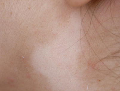
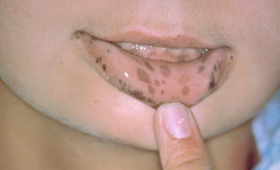
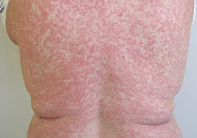
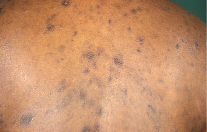
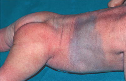
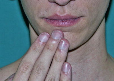
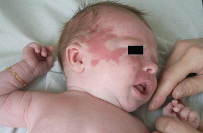
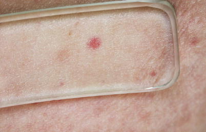

Fig. 3.1
White macule. Vitiligo. A macule is a localized anomaly of the color or transparency of the skin. By definition, a macule is not palpable. This example is a white macule observed in vitiligo. Vitiligo causes depigmentation of the skin. This autoimmune disorder of unknown origin can sometimes be associated with other autoimmune disorders such as thyroiditis or Biermer’s disease. Note that the vellus hair present on the leukodermic macule in this patient is also depigmented

Fig. 3.2
Pigmented macules. Lentigo. Multiple brown macules of the lips. This is a case of mucosal lentigo. When macules are numerous and localized on the lips, they can be markers for Peutz-Jeghers syndrome, a diagnosis that should then be considered (see Chap. 15). In this syndrome, lentigines are associated with gastrointestinal polyposis as well as an increased risk of some non-gastrointestinal cancers

Fig. 3.3
Maculopapular exanthema. Skin reaction after use of dextropropoxyphene. Exanthema indicates the rapid and eruptive onset of circumscribed skin lesions on almost the entire integument. The lesions may be confluent, as shown in this example. These are macules, papules, and red plaques. In this example, the eruption was due to an analgesic allergy (“cutaneous drug reaction”). This type of drug reaction is common (see Chap. 12). Lesions disappear within a few days after discontinuation of the drug

Fig. 3.4
Postinflammatory macules of the back. Lichen (planus). Brown pigmented macules of strong or mild coloration, resulting from lichen. According to the phototype of the individual, many inflammatory dermatoses can leave temporary or longer-lasting sequelae which are either hypopigmented (“postinflammatory hypopigmentation”) or hyperpigmented (“postinflammatory hyperpigmentation”). Because they do not have any histopathological specificity, a retrospective diagnosis cannot be made

Fig. 3.5
Cerulodermic macule. Mongolian spot. Extended bluish-gray macule of several centimeters in a newborn. These lesions, called Mongolian spots, are common among newborns of Asian and North African origin. They have no special implication and usually regress with time

Fig. 3.6
Cyanosis in the context of hepatopulmonary syndrome. Bluish aspect of the lips and extremities, typical of a central cyanosis. Central cyanosis reflects a significant reduction in the arterial oxygen saturation (at least 2–3 g deoxyhemoglobin/100 mL), whereas peripheral cyanosis is the result of excessive extraction of oxygen in peripheral tissues, thus explaining its location at the extremities and the nose and the sparing of mucous membranes

Fig. 3.7
Angiomatous macule. Capillary malformation (“port-wine stain”). Dark red “angiomatous” macules systematically distributed on V2 territory, in a port-wine stain (nevus flammeus). This type of angioma is not rare. When segmentally distributed, as is here on the territory of one of the branches of the trigeminal nerve, it may be the expression of a cutaneomeningospinal angiomatosis in the context of Sturge-Weber syndrome. This is especially true if sited on the V1 territory

Fig. 3.8
Purpuric macule. Diascopy. Red macule not disappearing when pressure is applied on the skin with a transparent glass forcing out blood from the vessels (diascopy technique): It is therefore purpura, resulting from an intracutaneous hemorrhage; red blood cells being no longer in the vessels, the color persists on diascopy
3.2 Erythema
Erythema is a localized or diffuse redness (Fig. 3.9) of the skin which disappears on diascopy. It is either permanent or paroxysmal, sometimes reticulated (livedo) (Fig. 3.10) and at other times bluish (erythrocyanosis). Its color varies from pale pink to dark red. Erythema is often associated with desquamation, thus producing erythematosquamous lesions (see Fig. 2.7).
Stay updated, free articles. Join our Telegram channel

Full access? Get Clinical Tree


