(1)
Hôpital Universitaire de Strasbourg, Strasbourg, France
13.1.1 Seborrheic Keratosis
13.1.2 Nevus
13.1.3 Vascular Lesions
13.1.4 Cysts
13.1.5 Other Lesions
13.2.1 Melanoma
13.2.2 Basal Cell Carcinoma
13.2.3 Spinous Cell Carcinoma
Abstract
Most individuals reaching adulthood are likely to develop a cutaneous tumor during their lifetime. Most of these tumors are benign. Some of them are malignant, either because of their ability to grow indefinitely and to invade local tissues, thus causing death (e.g., basal cell carcinoma), or because of their metastatic ability to spread (e.g., melanoma). For teaching purposes, this chapter presents certain lesions which are not tumors in the strict sense, but can suggest the possibility of this diagnosis (e.g., stucco keratosis, venous lake).
Most individuals reaching adulthood are likely to develop a cutaneous tumor during their lifetime. Most of these tumors are benign. Some of them are malignant, either because of their ability to grow indefinitely and to invade local tissues, thus causing death (e.g., basal cell carcinoma), or because of their metastatic ability to spread (e.g., melanoma). For teaching purposes, this chapter presents certain lesions which are not tumors in the strict sense, but can suggest the possibility of this diagnosis (e.g., stucco keratosis, venous lake).
13.1 Tumors and Benign Proliferations
13.1.1 Seborrheic Keratosis
Papules and brown keratotic plaques (Fig. 13.1). Seborrheic keratosis is a very common benign tumor, particularly in patients over 60 years old. Most seborrheic keratoses are pigmented, which calls for differential diagnosis with nevi and melanomas. Since this type of tumor is so common, every physician should be able to recognize it, at least in its typical form. It is a proliferation of epidermal cells with characteristic clinical appearances such as papules, plaques, or nodules that are generally keratotic, with sharp demarcations and rectangular borders (lesion appears as if laid on the skin), roughness on palpation, horny plugs (intralesional keratoses that originate from dilated ostia of hair follicles), and color ranging from light brown to dark brown, and are sometimes heterochromatic (Fig. 13.2). In Castellani’s disease, numerous brown keratotic papules similar to those found in seborrheic keratosis can be observed on dark skin, in the facio-cervical areas and the upper trunk (also called dermatosis papulosa nigra, Fig. 13.3). When seborrheic keratoses are eruptive, multiple, and inflammatory, they can be the manifestation of a paraneoplastic syndrome (Leser-Trélat sign).
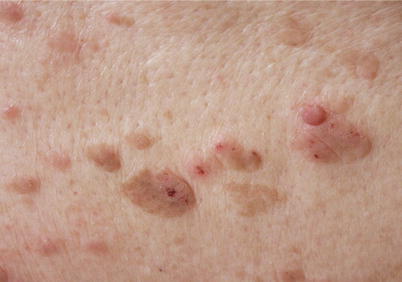
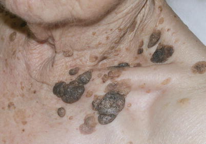
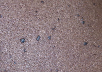
Fig. 13.1
∎
Fig. 13.2
∎
Fig. 13.3
∎
13.1.2 Nevus
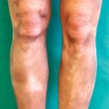
Hemispheric, brown, and dome-shaped papule of slightly less than 1 cm wide (Fig. 13.4). A nevus is commonly referred to as a mole. This type of tumor is very common on white skin (Fig. 13.5). It can be a macule, a papule, or a nodule. The size and shape are variable (Fig. 13.6). Color ranges from light brown to black; there are also bluish variants (Fig. 13.7) and even achromic nevi. The surface may be multilobulated (or mammillate) (Fig. 13.4). A nevus can be acquired or congenital. Congenital lesions are often large (Fig. 13.8). A white ring may surround one or more nevi (Sutton’s phenomenon or Sutton’s nevi); some of these nevi will thus involute and become achromic (Fig. 13.9). The occurrence of a Sutton’s phenomenon commands a thorough examination, since it may be synchronous to an evolving melanoma. When several nevi are affected (Sutton’s disease, Fig. 13.9), it can also be a forewarning sign of vitiligo.
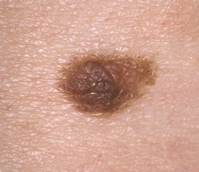
Fig. 13.4
∎
Characteristics such as symmetry, evenness, and absence of evolutivity allow distinguishing a nevus from a melanoma.
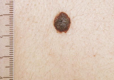
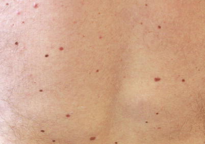
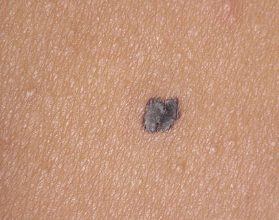
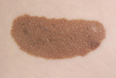
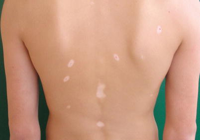
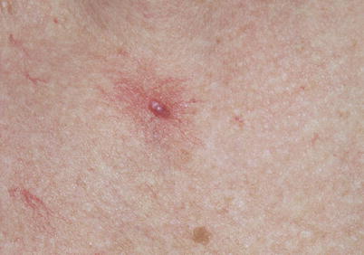
Fig. 13.5
∎
Fig. 13.6
∎
Fig. 13.7
∎
Fig. 13.8
∎
Fig. 13.9
∎
Fig. 13.10
∎
13.1.3 Vascular Lesions
Red telangiectatic spot with an angiomatous papule in its center. Spider nevus (or nevus araneus) (Fig. 13.10). Pressure exerted on the central papule “empties” the lesion, which reappears by centripetal filling, after pressure is taken off. This type of angioma is quite common; it can appear during pregnancy and disappear after delivery. Multiple spider nevi may indicate a state of relative hyperestrogenism and be one of the cutaneous signs of hepatic cirrhosis.
Reddish-black papule. Thrombosed angioma (Fig. 13.11). Note the peripheral, blue-black, and even spots. This type of lesion is sometimes clinically mistaken for a melanoma. However, anamnesis will find the notion that there has been a preceding red lesion which has changed rapidly over a few days and become painful. A biopsy is indispensable in case of doubt.
Fleshy and erosive nodule, partially covered with epidermis (epidermalized), that complicates an ingrown nail (Fig. 13.12). Botryomycoma (also known as pyogenic granuloma or proud flesh). Proud flesh (Fig. 13.13) usually occurs rapidly. These lesions may resemble achromic melanoma, especially when located in extremities (cf. Fig. 13.29).
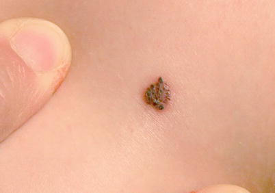
Fig. 13.11


∎
Related posts:

Stay updated, free articles. Join our Telegram channel
Full access? Get Clinical Tree
