3
Fever and Rash
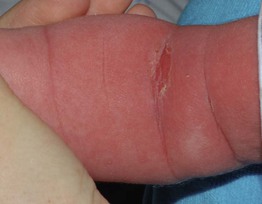
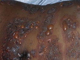
• A variety of infectious and inflammatory conditions can present with fever and a rash (Fig. 3.1). The cutaneous findings range from a morbilliform eruption or urticaria (Figs. 3.2 and 3.3) to confluent erythema (Fig. 3.4) to petechial, vesiculobullous, and pustular lesions (Figs. 3.5–3.9).
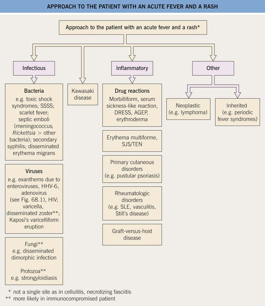
Fig. 3.1 Approach to the patient with an acute fever and a rash. AGEP, acute generalized exanthematous pustulosis; DRESS, drug reaction with eosinophilia and systemic symptoms (also referred to as drug-induced hypersensitivity syndrome [DIHS]); HHV, human herpes virus; HIV, human immunodeficiency virus; SJS, Stevens–Johnson syndrome; SLE, systemic lupus erythematosus; SSSS, staphylococcal scalded skin syndrome; TEN, toxic epidermal necrolysis.
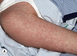
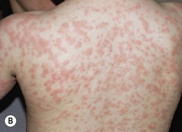
Fig. 3.2 Morbilliform drug eruptions. A Fine pink macules and thin papules, becoming confluent on the posterior upper arm, which is a dependent area in this hospitalized patient. B More edematous (‘urticarial’) pink papules; unlike true urticaria, these lesions are not transient. Courtesy, Julie V. Schaffer, MD.
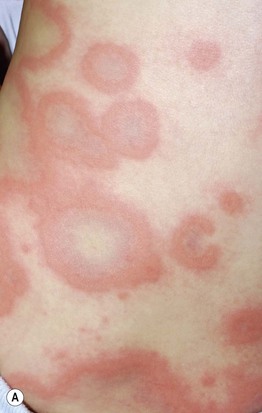
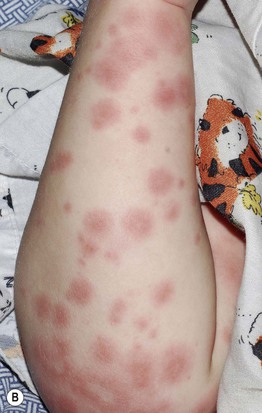
Fig. 3.3 Urticaria and serum sickness-like reaction. A Giant annular urticaria (urticaria ‘multiforme’) in a young child with a recent viral upper respiratory tract infection. Individual lesions last <24 hours, but they often resolve with a dusky purplish hue that can lead to misdiagnosis as erythema multiforme. B Serum sickness-like reaction due to amoxicillin. Some of the urticarial papules and annular plaques have a purpuric component, and the eruption was accompanied by high fevers, lymphadenopathy, arthralgias, and acral edema. Courtesy, Julie V. Schaffer, MD.
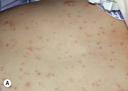
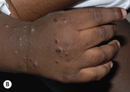
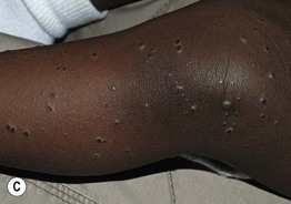
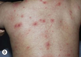
Fig. 3.5 Viral exanthems. A Enteroviral exanthem presenting as widespread small pink papules, many with petechiae centrally. B, C Widespread vesicular eruption due to coxsackievirus A6 infection. D Scattered vesicles on erythematous bases in varicella, with lesions in different stages of evolution. Courtesy, Julie V. Schaffer, MD.
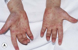
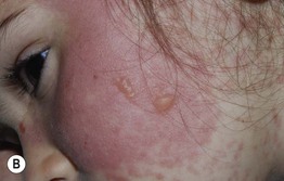
Fig. 3.6 Stevens–Johnson syndrome. The patient initially developed multiple small pink papules mimicking a morbilliform eruption, but with accentuation on the palms (A). A day later, confluent erythema and bullae had developed (B), and involvement of the vermilion lips and conjunctiva was evident. Courtesy, Julie V. Schaffer, MD.
