and Peter M. Prendergast2
(1)
Elysium Aesthetics, Bogota, Colombia
(2)
Venus Medical, Dublin, Ireland
Introduction
Beautiful, slim, and shapely female legs are synonymous with sex appeal. Although long, toned, or slightly muscular women’s legs might be generally perceived to be attractive, there is little consensus of what exact shapes and contours an attractive leg should have (Fig. 18.1). The curves, shapes, and spaces between the legs all contribute to the aesthetics of the lower limb.
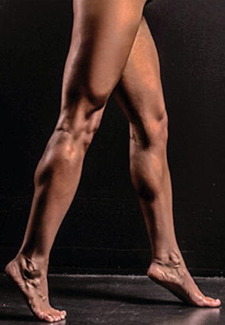

Fig. 18.1
Athletic female legs
Since the lower extremity is an area of high exposure in females, body contouring requires precision because there is little room for error. One of the challenges is that there are multiple adhesion zones and danger areas for liposuction in the lower limb [1, 2].
Some surgeons do not perform liposculpture of the legs due to artistic and/or technical difficulties. There is a scarcity of published medical data pertaining to beauty standards, aesthetic ideals, and surgical techniques for the lower limb [3]. We are now describing a form of ultrasound-assisted lipoplasty that represents a safe, effective, and reproducible technique for enhanced definition of the lower limb.
The Ideal Leg
The author describes a simple method to delineate the perfect leg following a series of convexities and concavities. Anterior and posterior contours, as well as medial and lateral ones are defined by muscular shape and volume, localized fat pads and bony promimences. All these components in balance define atractive legs.
Medial
Starting proximally, there should be a visible “rhomboid of light” between the upper inner thighs. The superior borders of the rhomboid are formed by the most medial aspect of the gluteal crease; the inferior borders by the inner thigh. Moving distally, the inner thigh forms a slight convexity, followed by a subtle concavity in the middle third. This concavity defines an adhesion zone and an area prone to contour irregularities. Below the zone of adherence, the lower limb has a gentle convex-concave-convex curvature that the author refers to as the double “S” shape: convex due to the femur and the knee fat pad, concave in the proximal calf, and convex over the medial gastrocnemius.
The distal calf has a slight concave line following the gastrocnemius shape toward the calcaneus bone.
Lateral
An important part of the female silhouette is formed by the smooth convex transition between the hip fat and the lateral thigh. Ideally, the trochanteric depression should not be visible in the female patient. The lateral thigh contour follows the shape of the quadriceps muscle and continues distally as a slight concavity, marking the site of the zone of adherence.
At the knee there is a continuation of the concavity and smooth transition to the proximal convexity of the leg over the lateral gastrocnemius, creating a “C” curve, followed by a subtle concavity distally towards the heel (Fig. 18.2).
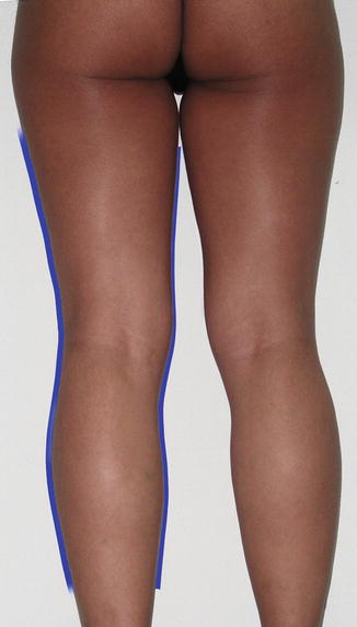

Fig. 18.2
Aesthetically ideal leg: good skin tone, balanced muscles in volume and shape, smooth curvilinear silhouette
Stealth Incisions
The legs play an important role in female physical attractiveness. Since they are regularly exposed, the incisions need to be as concealed as possible. Moreover, the neurovascular anatomy in the lower limb forbids us to make access incisions in certain areas [4–6]. The incisions for lower limb liposculpture are:
1.
Infragluteal crease midpoint (IGM): This incision provides access to the lower half of the buttocks, posterior and inner thigh, banana roll, and lateral thigh.
2.
Pubic incision: This allows access to the anterior leg and the inner anterior thigh. With the use of long cannulae, the lower thigh can also be reached.
3.
Knee incision: An incision over the upper portion of the patella provides access to the lower leg, medial thigh, medial knee fat pad, and the lateral thigh.
4.
Posteromedial knee point (PMK): This incision provides access to the middle, lower, and medial posterior thigh as well as the upper portion of the posterior and medial calf. Do not cross to the lateral side from this site to avoid neurovascular injury [5].
5.
Posterolateral knee point (PLK): From this incision we can access the middle and lower lateral thigh as well as the lateral and posterior calf. Again, to avoid neurovascular injury, avoid crossing to the medial side form this access incision [6]. Special care should be taken to avoid the common peroneal nerve [4].
6.
Achilles tendon incision: An incision in the skin over the tendon provides access to the medial and lateral aspects of the lower and middle calf. The incision should be placed above the level of the superior borders of the malleoli to avoid inadvertent injury to vascular and neural structures (Fig. 18.3) [6].
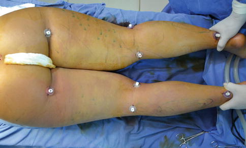

Fig. 18.3
Posterior lower limb incisions with ports
The Use of Drains
Drains are not necessary for thighs or calves, but lower incisions can be left open for permissive drainage. Drainage through the PMK and PLK incisions is facilitated by gravity and external compression.
Markings
Deep Markings
The general markings will start from proximally in zones 3 and 4 in the subgluteal area (Fig. 18.4).
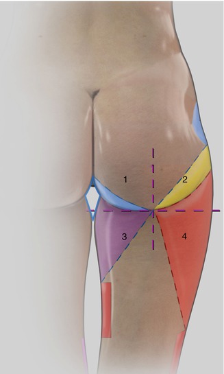

Fig. 18.4
Inferior perigluteal and leg zones: 1 lower internal angle (blue); 2 transition zone between the lateral thigh and the external gluteal fold (yellow); 3 inner thigh (purple); 4 lateral thigh (red)
Zone 3 is marked to delineate the upper inner thigh fat. The markings extend inferiorly over the middle third of the inner thigh so that there is a smooth transition form the slight convexity of the upper inner thigh to the flattened or slight concave area at the zone of adherence.
Zone 4, the outer thigh, extends from the lateral portion of the buttocks (described in Chap. 15) to the inferior limit of the trochanteric depression superiorly and inferiorly to the distal third of the lateral thigh where there is an adhesion zone (Fig. 18.5). This zone requires deep liposuction to debulk the area that deforms the hips. There should be a smooth convex lateral curve from the waist to the distal third of the lateral thigh, without interruption by the trochanteric depression.
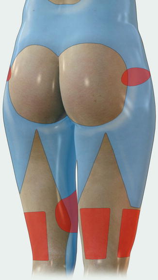

Fig. 18.5
Adhesion zones in the lower limb: trochanteric, distal third of lateral and posterior thigh, and the middle-third of the medial thigh


