Facial Transplantation— The Anatomic Basis
M. SIEMIONOW
G. AGAOGLU
S. UNAL
EDITORIAL COMMENT
Increasing numbers o composite allotransplantation of the face are being performed throughout the world. Anyone who is interested in facial transplantation will be well served by studying the descriptive anatomy of the donor and the recipient dissections as outlined in this chapter.
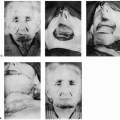
Lifelong immunosuppression therapy, due to serious side effects, is the main obstacle to routine use of composite tissue allografts. When the subject is a candidate for face transplantation, there are ethical, social, and psychologic issues to be considered. In addition, the technical aspects of face transplantation have to be outlined. To mimic the clinical scenario of a facial transplantation procedure, we have performed a mock facial transplantation by harvesting a total facial-scalp flap from donor cadavers and transferring the flap to recipient cadavers.
INDICATIONS
Since the first successful hand allograft transplantation in France in 1998 (1), composite tissue allograft transplantation has opened a new era in the field of reconstructive surgery. However, the final cosmetic and functional outcomes of currently available reconstructive procedures for severe soft-tissue defects of the face are not optimal. Face transplantation is a promising alternative to the conventional reconstructive procedures. Successful partial face transplantation cases have been reported in France and in China (2, 3, 4). We have designed a cadaveric facial allograft transplantation model (5, 6) to support application of facial allografts, as a new treatment option for reconstruction of facial defects in patients with severe facial deformities due to burn, trauma, and cancer ablation.
ANATOMY
In donor cadavers, a full facial-scalp flap was elevated to include the entire facial skin and the scalp. The flap also incorporated the superficial muscular aponeurotic system (SMAS) of the face; the parotid gland; the inferior and superior tarsal plates; and the great auricular, facial, supraorbital, infraorbital, and mental nerves. This composite facial-scalp flap is based bilaterally on the external carotid arteries, including superficial temporal and facial arteries, and on the external jugular and facial veins.
FLAP DESIGN AND DIMENSIONS
The flap was designed to include the entire facial skin and the scalp, including both ears, based on the external carotid arteries and external jugular and facial veins. The mean horizontal-vertical dimensions for the total facial-scalp flaps were 57 × 30 cm, and the mean surface area was 1,192 cm2. The mean horizontal and vertical dimensions for facial flaps without scalp were 33 × 28 cm, and the mean surface area was 675 cm2.
OPERATIVE TECHNIQUE
Mock Facial Transplantation: Technique of Donor Facial-Scalp Flap Harvesting
A midline vertical incision from mentum to suprasternal notch was performed to the depth of the platysmal layer (Fig. 151.1A). From the lower end of the vertical incision, horizontal incisions were extended bilaterally, to be met posteriorly in the neck midline at the scalp hairline. With the cadaver in the left lateral position, the incision was extended vertically in the midline to the vertex of the scalp.
In the subplatysmal plane, the flaps were elevated from medial to lateral and caudal to cranial, exposing the strap muscles at the midline and the sternocleidomastoid muscle (SCM) more laterally. The caudal portions of the external jugular
veins (EJVs) were found bilaterally underneath the platysmal layer, ligated, transected, and incorporated into the skin-platysmal flap. The great auricular nerves were both identified on the lateral edges of the SCM and incorporated into the flap.
veins (EJVs) were found bilaterally underneath the platysmal layer, ligated, transected, and incorporated into the skin-platysmal flap. The great auricular nerves were both identified on the lateral edges of the SCM and incorporated into the flap.
The scalp-flap dissection was performed in the subgaleal plane, with the cadaver turned to the left lateral and right lateral positions. The supraorbital rims were reached, and the supraorbital nerves were traced down to their exit from the supraorbital foramen and transected. Then, the external ear canals were reached and transected to incorporate the ears within the flap.
In the neck, the SCM was cut close to its clavicular and sternal insertions, and the carotid sheath underneath was exposed. The omohyoid muscle was cut at its tendinous junction, to better expose the caudal portion of the carotid sheath. The carotid sheath was incised and the carotid artery, along with the internal jugular vein, was dissected cranially to reach the carotid bifurcation. After the bifurcation of the common carotid to internal and external carotid arteries, the external carotid artery was transected, to become the arterial pedicle of the flap.
At this point, the attachments of the stylohyoid and digastric muscles on the hyoid bone were cut, to get a better exposure of the underlying structures. The ascending pharyngeal, the superior thyroid, and the lingual arteries were ligated close to their branching from the external carotid, and the dissection was further carried cranially. The hypoglossal nerve was also exposed and transected. The facial artery, branching from the anteromedial side of the external carotid artery, was carefully dissected in a cranial direction as it entered the submandibular gland. The artery was traced and separated from the gland with a tedious dissection, and the gland was not incorporated in the platysmal skin flap (Fig. 151.1B).
Then, the second venous pedicle of the flap, the facial vein, was also isolated from the surrounding soft tissue and the submandibular gland. After dissection of the vein toward its entrance to the internal jugular vein, it was ligated and transected, leaving sufficient pedicle to become the second venous pedicle of the flap. After the mandibular border was reached at the midline, the dissection was continued cranially underneath the SMAS, to reach the lips. The vermilion of the lips was incorporated within the flap, as the sub-SMAS dissection was deepened to incise the gingivobuccal mucosa. Through intraoral incision, the mental nerves were exposed, traced, and incorporated within the flap.
After keeping the facial artery intact, the external carotid artery was further traced cranially behind the ramus of the mandible as it continued deep to the parotid gland. The gland was incorporated within the flap. Before becoming the superficial temporal artery, the external carotid gives off the occipital, the maxillary, the transverse facial, and the middle temporal branches, each of which was ligated and transected sequentially.
In the cheek flap dissections continued in a sub-SMAS plane, a fashion similar to a face-lift dissection was used. Intraoral incision was circumferentially carried onto the upper gingivobuccal sulcus, and the soft tissues were sharply incised. Through the upper gingivobuccal incision, in the subperiosteal plane, the infraorbital nerve was explored, traced, and included in the flap.
On both sides, the upper and lower palpebral fornix conjunctival incisions were performed in a circumferential manner just at the tarsal margins and joined with the sub-SMAS flap dissection plane, in order to incorporate the upper and lower lids within the flap. At the midline, the nasal soft tissue was elevated, leaving the osseocartilaginous framework behind, and the elevation of the total facial-scalp flap was completed (Fig. 151.1C and D).
Technique of Recipient Facial Skin Harvesting as a “Monoblock” Full-Thickness Skin Graft
Incisions were made in a fashion similar to that described previously. The entire skin of the neck, face, and scalp was incised and elevated as a “monoblock” full-thickness skin graft in a plane above the platysma, SMAS, and galea, respectively (Fig. 151.2A and B). The great auricular nerve was identified at the posterior border of the SCM muscle, dissected from the surrounding tissue, and the maximal access to the nerve was preserved. Through the upper and lower gingivobuccal incisions, the vermilion of the upper and lower lips was excised with the entire facial skin. At the infraorbital and mental regions, both the infraorbital and mental nerves were explored and dissected from their surrounding tissue, respectively, until an adequate length of the nerves was achieved for future coaptation. The upper and lower circumferential conjunctival incisions were performed at the tarsal margins and were joined with the facial skin dissection plane, incorporating the upper and lower lids. At the supraorbital rims, the supraorbital nerves were explored and traced down to the supraorbital foramen, until adequate length was obtained.
Next, the arterial and venous pedicles were dissected and prepared for the future anastomoses. External jugular veins were bilaterally explored under the platysma and were prepared to serve as one of the recipient venous pedicles. SCM muscle was retracted laterally, and the carotid sheath was reached and incised. The external carotid artery was dissected, separated from the internal carotid artery, and prepared as the recipient’s arterial pedicle. The facial vein was identified at its entrance into the internal jugular vein and dissected to obtain adequate length to serve as the second venous pedicle in the recipient.
Related posts:


Stay updated, free articles. Join our Telegram channel

Full access? Get Clinical Tree


